Attenuation of salt-induced cardiac remodeling and diastolic dysfunction by the GPER agonist G-1 in female mRen2.Lewis rats
- PMID: 21082029
- PMCID: PMC2972725
- DOI: 10.1371/journal.pone.0015433
Attenuation of salt-induced cardiac remodeling and diastolic dysfunction by the GPER agonist G-1 in female mRen2.Lewis rats
Abstract
Introduction: The G protein-coupled estrogen receptor (GPER) is expressed in various tissues including the heart. Since the mRen2.Lewis strain exhibits salt-dependent hypertension and early diastolic dysfunction, we assessed the effects of the GPER agonist (G-1, 40 nmol/kg/hr for 14 days) or vehicle (VEH, DMSO/EtOH) on cardiac function and structure.
Methods: Intact female mRen2.Lewis rats were fed a normal salt (0.5% sodium; NS) diet or a high salt (4% sodium; HS) diet for 10 weeks beginning at 5 weeks of age.
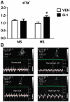
Results: Prolonged intake of HS in mRen2.Lewis females resulted in significantly increased blood pressure, mildly reduced systolic function, and left ventricular (LV) diastolic compliance (as signified by a reduced E deceleration time and E deceleration slope), increased relative wall thickness, myocyte size, and mid-myocardial interstitial and perivascular fibrosis. G-1 administration attenuated wall thickness and myocyte hypertrophy, with nominal effects on blood pressure, LV systolic function, LV compliance and cardiac fibrosis in the HS group. G-1 treatment significantly increased LV lusitropy [early mitral annular descent (e')] independent of prevailing salt, and improved the e'/a' ratio in HS versus NS rats (P<0.05) as determined by tissue Doppler.
Conclusion: Activation of GPER improved myocardial relaxation in the hypertensive female mRen2.Lewis rat and reduced cardiac myocyte hypertrophy and wall thickness in those rats fed a high salt diet. Moreover, these advantageous effects of the GPER agonist on ventricular lusitropy and remodeling do not appear to be associated with overt changes in blood pressure.
Conflict of interest statement
Figures


References
-
- Kitzman DW, Gardin JM, Gottdiener JS, Arnold A, Boineau R, et al. Importance of heart failure with preserved systolic function in patients > or = 65 years of age. CHS Research Group. Cardiovascular Health Study. Am J Cardiol. 2001;87:413–419. - PubMed
-
- Masoudi FA, Havranek EP, Smith G, Fish RH, Steiner JF, et al. Gender, age, and heart failure with preserved left ventricular systolic function. J Am Coll Cardiol. 2003;41:217–223. - PubMed
-
- Solomon SD, Verma A, Desai A, Hassanein A, Izzo J, et al. Effect of intensive versus standard blood pressure lowering on diastolic function in patients with uncontrolled hypertension and diastolic dysfunction. Hypertension. 2010;55:241–248. - PubMed
-
- Weinberger MH. Salt sensitivity as a predictor of hypertension. Am J Hypertens. 1991;4:615S–616S. - PubMed
-
- Heimann JC, Drumond S, Alves AT, Barbato AJ, Dichtchekenian V, et al. Left ventricular hypertrophy is more marked in salt-sensitive than in salt-resistant hypertensive patients. J Cardiovasc Pharmacol. 1991;17(Suppl 2):S122–S124. - PubMed
Publication types
MeSH terms
Substances
Grants and funding
LinkOut - more resources
Full Text Sources
Other Literature Sources
Medical
Miscellaneous

