

Capillary hemangioma of the thoracic spinal cord
- PMID: 21082058
- PMCID: PMC2966732
- DOI: 10.3340/jkns.2010.48.3.272
Capillary hemangioma of the thoracic spinal cord
Abstract
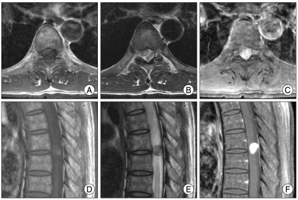
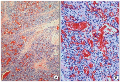
Capillary hemangiomas are common soft tissue tumors on the skin or mucosa of the head and neck in the early childhood, but very rare in the neuraxis. A 47-year-old man presented with one month history of back pain on the lower thoracic area, radiating pain to both legs, and hypesthesia below T7 dermatome. Thoracic spine MRI showed 1×1.3×1.5 cm, well-defined intradural mass at T6-7 disc space level, which showed isointensity to spinal cord on T1, heterogeneous isointensity on T2-weighted images, and homogeneous strong enhancement. The patient underwent T6-7 total laminotomy, complete tumor removal and laminoplasty. Histologically, the mass showed a capsulated nodular lesion composed of capillary-sized vascular channels, which were tightly packed into nodules separated by fibrous septa. These features were consistent with capillary hemangioma.
Keywords: Capillary hemangioma; Intradural extramedullary tumor; Spinal cord.
Figures
Similar articles
-
Intradural extramedullary capillary hemangioma in the upper thoracic spine: a review of the literature.Case Rep Orthop. 2014;2014:604131. doi: 10.1155/2014/604131. Epub 2014 Jun 18. Case Rep Orthop. 2014. PMID: 25045565 Free PMC article.
-
Spinal intradural capillary hemangioma.Surg Neurol. 2006 Aug;66(2):212-4. doi: 10.1016/j.surneu.2005.11.018. Surg Neurol. 2006. PMID: 16876637
-
Intradural extramedullary capillary hemangioma of lower thoracic spinal cord.Indian J Orthop. 2012 Jul;46(4):475-8. doi: 10.4103/0019-5413.97262. Indian J Orthop. 2012. PMID: 22912525 Free PMC article.
-
Spinal epidural capillary hemangioma: A systematic literature review and an illustrative case.Neurochirurgie. 2022 Dec;68(6):697-701. doi: 10.1016/j.neuchi.2022.03.004. Epub 2022 Apr 25. Neurochirurgie. 2022. PMID: 35477014
-
[Capillary hemangioma of the spinal cord. A new case].Neurochirurgie. 2002 Nov;48(5):440-4. Neurochirurgie. 2002. PMID: 12483124 Review. French.
Cited by
-
Intradural extramedullary capillary hemangioma of the cauda equina: Case report and literature review.Surg Neurol Int. 2015 Apr 22;6(Suppl 3):S127-31. doi: 10.4103/2152-7806.155701. eCollection 2015. Surg Neurol Int. 2015. PMID: 25949855 Free PMC article.
-
Spinal Intradural Extramedullary Capillary Hemangioma with Coexistent Spinal Edema and Syringomyelia Successfully Treated by Tumor Removal and Cervical Laminoplasty.Asian J Neurosurg. 2021 Dec 18;16(4):854-871. doi: 10.4103/ajns.AJNS_51_21. eCollection 2021 Oct-Dec. Asian J Neurosurg. 2021. PMID: 35071092 Free PMC article.
-
A case report of lobular intradural extramedullary capillary hemangioma in a 14-year-old patient: resection and reconstruction.J Spine Surg. 2024 Mar 20;10(1):152-158. doi: 10.21037/jss-23-113. Epub 2024 Mar 14. J Spine Surg. 2024. PMID: 38567015 Free PMC article.
-
Spinal capillary hemangiomas: Two cases reports and review of the literature.Asian J Neurosurg. 2017 Jul-Sep;12(3):556-562. doi: 10.4103/1793-5482.148793. Asian J Neurosurg. 2017. PMID: 28761543 Free PMC article.
-
Rapidly Progressive Atypical Vertebral Hemangioma: A Case Report.Korean J Neurotrauma. 2020 Aug 20;16(2):320-325. doi: 10.13004/kjnt.2020.16.e24. eCollection 2020 Oct. Korean J Neurotrauma. 2020. PMID: 33163444 Free PMC article.
References
-
- Abe M, Tabuchi K, Tanaka S, Hodozuka A, Kunishio K, Kubo N, et al. Capillary hemangioma of the central nervous system. J Neurosurg. 2004;101:73–81. - PubMed
-
- Abe M, Misago N, Tanaka S, Masuoka J, Tabuchi K. Capillary hemangioma of the central nervous system : a comparative study with lobular capillary hemangioma of the skin. Acta Neuropathol. 2005;109:151–158. - PubMed
-
- Enzinger FM, Weiss SW. Benign tumors and tumor-like lesions of blood vessels in soft tissues tumors : soft tissue tumors. ed 3. St. Louis: Mosby; 1995. pp. 579–626.
-
- Hanakita J, Suwa H, Nagayasu S, Suzuki H. Capillary hemangioma in the cauda equina : neuroradiological findings. Neuroradiology. 1991;33:458–461. - PubMed
Publication types
LinkOut - more resources
Full Text Sources
