

Neutralization of BDNF attenuates the in vitro protective effects of olfactory ensheathing cell-conditioned medium on scratch-insulted retinal ganglion cells
- PMID: 21082236
- PMCID: PMC11498652
- DOI: 10.1007/s10571-010-9626-5
Neutralization of BDNF attenuates the in vitro protective effects of olfactory ensheathing cell-conditioned medium on scratch-insulted retinal ganglion cells
Abstract
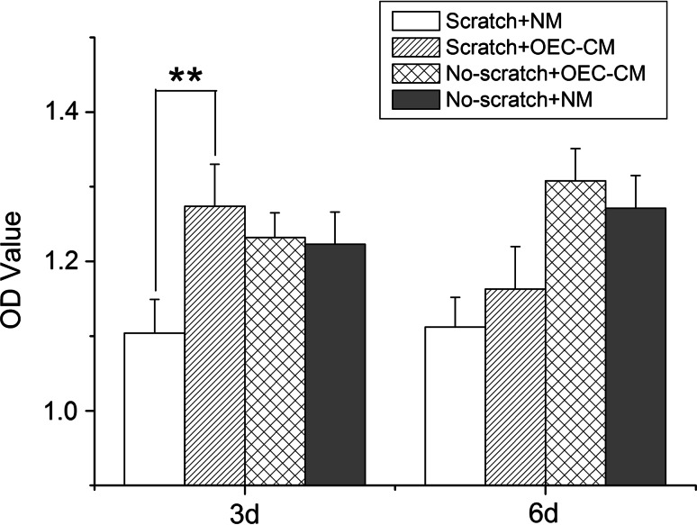
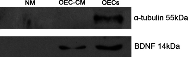
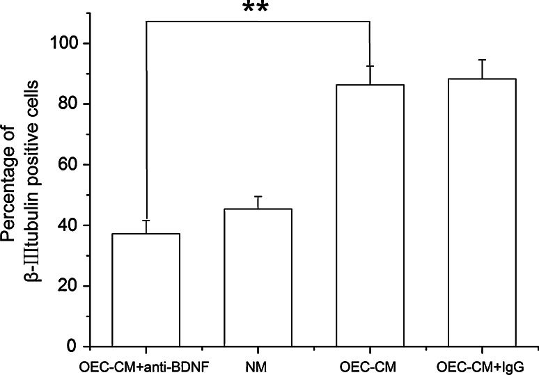
Transplantation of olfactory ensheathing cells (OECs) becomes one of the promising strategies in restoring lost functions of injured central nervous system. Elevated level of expressed brain-derived neurotrophic factor (BDNF) was revealed in the previous studies to be related to the protective effects of OECs on injured cortical and brain stem neurons as well as retinal ganglion cells (RGCs), but no evidence has been obtained to demonstrate whether transplanted OECs protect injured central neurons directly by their secreted BDNF. In the present study, the effects of BDNF neutralization on the neuroprotection of adult OEC-conditioned medium (OEC-CM) on scratch-insulted RGCs were examined. The results showed that OEC-CM protected cultured RGCs from scratch insult, and neutralization of BDNF by BDNF neutralizing antibody attenuated such neuroprotection of the medium. It is thus concluded that neurotrophic factors including BDNF secreted by OECs can protect injured OECs in vitro and BDNF plays a major role in such a protection of OECs.
Figures
Similar articles
-
Brain-derived Neurotrophic Factor Promotes the Migration of Olfactory Ensheathing Cells Through TRPC Channels.Glia. 2016 Dec;64(12):2154-2165. doi: 10.1002/glia.23049. Epub 2016 Aug 18. Glia. 2016. PMID: 27534509
-
The differentiation of rat adipose-derived stem cells into OEC-like cells on collagen scaffolds by co-culturing with OECs.Neurosci Lett. 2007 Jun 29;421(3):191-6. doi: 10.1016/j.neulet.2007.04.081. Epub 2007 Jun 2. Neurosci Lett. 2007. PMID: 17574753
-
Transplanted olfactory ensheathing cells reduce retinal degeneration in Royal College of Surgeons rats.Curr Eye Res. 2012 Aug;37(8):749-58. doi: 10.3109/02713683.2012.697972. Epub 2012 Jun 12. Curr Eye Res. 2012. PMID: 22691022
-
[Olfactory ensheathing cells promote the survival of newborn rat spiral ganglion cells in vitro].Sheng Li Xue Bao. 2010 Apr 25;62(2):115-21. Sheng Li Xue Bao. 2010. PMID: 20401445 Chinese.
-
BDNF production by olfactory ensheathing cells contributes to axonal regeneration of cultured adult CNS neurons.Neurochem Int. 2007 Feb;50(3):491-8. doi: 10.1016/j.neuint.2006.10.004. Epub 2006 Dec 8. Neurochem Int. 2007. PMID: 17157963
Cited by
-
Delayed administration of bone marrow mesenchymal stem cell conditioned medium significantly improves outcome after retinal ischemia in rats.Invest Ophthalmol Vis Sci. 2014 Apr 3;55(6):3785-96. doi: 10.1167/iovs.13-11683. Invest Ophthalmol Vis Sci. 2014. PMID: 24699381 Free PMC article.
-
Conditioned medium of Wnt/β-catenin signaling-activated olfactory ensheathing cells promotes synaptogenesis and neurite growth in vitro.Cell Mol Neurobiol. 2013 Oct;33(7):983-90. doi: 10.1007/s10571-013-9966-z. Epub 2013 Jul 28. Cell Mol Neurobiol. 2013. PMID: 23893371 Free PMC article.
-
Enhancing the Therapeutic Potential of Olfactory Ensheathing Cells in Spinal Cord Repair Using Neurotrophins.Cell Transplant. 2018 Jun;27(6):867-878. doi: 10.1177/0963689718759472. Epub 2018 May 31. Cell Transplant. 2018. PMID: 29852748 Free PMC article. Review.
-
Extracellular Vesicles Derived From Olfactory Ensheathing Cells Promote Peripheral Nerve Regeneration in Rats.Front Cell Neurosci. 2019 Dec 6;13:548. doi: 10.3389/fncel.2019.00548. eCollection 2019. Front Cell Neurosci. 2019. PMID: 31866834 Free PMC article.
-
Dose-dependent protective effect of lithium chloride on retinal ganglion cells is interrelated with an upregulated intraretinal BDNF after optic nerve transection in adult rats.Int J Mol Sci. 2014 Aug 5;15(8):13550-63. doi: 10.3390/ijms150813550. Int J Mol Sci. 2014. PMID: 25100168 Free PMC article.
References
-
- Boruch AV, Conners JJ, Pipitone M, Deadwyler G, Devies GH, Jones KJ (2001) Neurotrophic and migratory properties of an olfactory ensheathing cell line. Glia 33(3):225–229 - PubMed
-
- Chen H, Weber AJ (2001) BDNF enhances retinal ganglion cell survival in cats with optic nerve damage. Invest Ophthalmol Vis Sci 42(5):966–974 - PubMed
-
- Chung RS, Adlard PA, Dittmann J, Vickers JC, Chuah MI, West AK (2004) Neuron-glia communication: metallothionein expression is specifically up-regulated by astrocytes in response to neuronal injury. J Neurochem 88(2):454–461 - PubMed
-
- Curtis R, Tonra JR, Stark JL, Adryan KM, Park JS, Cliffer KD, Lindsay RM, Distefano PS (1998) Neuronal injury increases retrograde axonal transport of the neurotrophins to spinal sensory neurons and motor neurons via multiple receptor mechanisms. Mol Cell Neurosci 12(3):105–118 - PubMed
Publication types
MeSH terms
Substances
LinkOut - more resources
Full Text Sources
