. 2011 Mar;31(2):323-35.
doi: 10.1007/s10571-010-9625-6. Epub 2010 Nov 17.

Immunocytochemical localization of TASK-3 (K(2P)9.1) channels in monoaminergic and cholinergic neurons

Affiliations

Immunocytochemical localization of TASK-3 (K(2P)9.1) channels in monoaminergic and cholinergic neurons

Christiane Marinc et al. Cell Mol Neurobiol. 2011 Mar.

Abstract

Monoaminergic and cholinergic systems are important regulators of cortical and subcortical systems, and a variety of vegetative functions are controlled by the respective neurotransmitters. Neuronal excitability and transmitter release of these neurons are strongly regulated by their potassium conductances carried by Kir and K(2P) channels. Here we describe the generation and characterization of a polyclonal monospecific antibody against rat TASK-3, a major brain K(2P) channel. After removal of cross-reactivities and affinity purification the antibody was characterized by ELISA, immunocytochemistry of TASK-3 transfected cells, and Western blots indicating that the antibody only detects TASK-3 protein, but not its paralogs TASK-1 and TASK-5. Western blot analysis of brain membrane fractions showed a single band around 45 kD, close to the predicted molecular weight of the TASK-3 protein. In addition, specific immunolabeling using the anti-TASK-3 antibody in Western blot analysis and immunocytochemistry was blocked in a concentration dependent manner by its cognate antigen only. Immunocytochemical analysis of rat brain revealed strong expression of TASK-3 channels in serotoninergic neurons of the dorsal and median raphe, noradrenergic neurons of the locus coeruleus, histaminergic neurons of the tuberomammillary nucleus and in the cholinergic neurons of the basal nucleus of Meynert. Immunofluorescence double-labeling experiments with appropriate marker enzymes confirmed the expression of TASK-3 in cholinergic, serotoninergic, and noradrenergic neurons. In the dopaminergic system strong TASK-3 expression was found in the ventral tegmental area, whereas TASK-3 immunoreactivity in the substantia nigra compacta was only weak. All immunocytochemical results were supported by in situ hybridization using TASK-3 specific riboprobes.


Figures
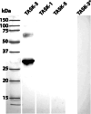
Fig. 1
Fig. 1
Western blot analysis of anti-TASK-3 antibody using recombinant TASK-1-, TASK-3-, and TASK-5-6HisTR fusion proteins. Gels were loaded with the respective fusion proteins (1 μg/lane). After electrophoresis and blotting the anti-TASK-3 antibody detected a single strong band around 28 kD, close to the predicted molecular weight of the TASK-3-6HisTR protein (6HisTR: thioredoxin with a 6 His N-terminal extension), and a weak band around 60 kD, presumably corresponding to a dimer. Preincubation of the anti-TASK-3 antibody with recombinant TASK-3-6HisTR protein (50 μg/ml, designated as TASK-3*) completely blocked TASK-3 specific immunostaining (outer right lane). Marker Dual color (Biorad) is indicated (outer left lane)
Fig. 2
Fig. 2
Characterization of monospecific anti-TASK-3 antibody by competitive ELISA. Microtitre plates were coated with the recombinant TASK-3-6HisTR antigen, the competitive antigens are indicated. Preincubation of the antibody with the cognate TASK-3-6HisTR decreased the signal in a concentration dependent manner, whereas TASK-1 and TASK-5 fusion proteins or GST had no effect
Fig. 3
Fig. 3
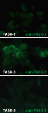
Immunoreactivity of transfected HT-22 cells. HT-22 cells transfected with TASK-3/pcDNA3.1 vector showed specific TASK-3 immunoreactivity. In constrast, TASK-1/pcDNA3.1 and TASK-5/pcDNA3.1 transfected cells were devoid of staining
Fig. 4
Fig. 4
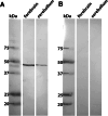
Western blot analysis of rat brain membrane fractions with the anti-TASK-3 antibody a Rat forebrain and cerebellar membrane fractions (up to 150 μg/lane) were separated by SDS-PAGE. After blotting only a single immunoreactive band around 45 kD is visible. b Preincubation of the anti-TASK-3 antibody with 50 μg/ml TASK-3-6HisTR antigen completely blocked specific immunostaining in both membrane fractions. Marker Dual color (Biorad) is indicated
Fig. 5
Fig. 5
Block of TASK-3 immunoreactivity by preincubation with TASK-3 antigen a Immunocytochemical analysis of a coronal section of the brain stem showed strong but not ubiquitous expression of TASK-3. Especially strong labeling was found in the dorsal raphe (inset). b Preincubation of the anti-TASK-3 antibody with 10 μg/ml TASK-3-GST antigen completely blocked specific immunostaining in the adjacent section. Bar represents 2 mm
Fig. 6
Fig. 6
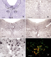
TASK-3 expression in serotoninergic dorsal and median raphe neurons a Cresyl violet staining of the mesencephalic raphe and surrounding nuclei (Bregma: −8.3 mm). c A considerable number of neurons in the dorsal raphe display TASK-3 immunoreactivity. e At higher magnification of c the localization of TASK-3 protein in somata and dendrites can be recognized. d The presence of TASK-3 mRNA as detected by in situ hybridization supports the expression of TASK-3 in DR neurons. f Double immunofluorescence discloses that the TASK-3 protein (Cy2, green) indeed is localized in serotonin-producing (Cy3, red) neurons (strong signal, yellow). b TASK-3 positive neurons are also found in median raphe neurons. DR, Dorsal raphe nucleus; MnR, median raphe nucleus; VTg, ventral tegmental nucleus; mlf, medial longitudinal fasciculus. Bar represent 1 mm in a, 200 μm in b, c, d, and 40 μm in e and f. (Color figure online)
Fig. 7
Fig. 7
TASK-3 channels are detected in dopaminergic neurons a Cresyl violet staining of midbrain region including the substantia nigra and the ventral tegmental area (Bregma: −5.3 mm). b Dopaminergic neurons of the SNC displayed medium but not strong TASK-3 immunoreactivity (arrows). c In contrast, strong TASK-3 expression was found in the VTA. d At higher magnification the localization of TASK-3 protein in somata and dendrites is appreciated. CG, Central gray; cp, cerebral peduncle; EW, Edinger-Westphal nucleus; IML, interstitial nucleus mlf; MG, medial geniculate nucleus; ml, medial lemniscus; mp, mammillary peduncle; RP, red nucleus parvocellular; SNC, substantia nigra pars compacta; SNR substantia nigra pars reticularis; VTA, ventral tegmental area. Bar represent 1 mm in a, 100 μm in b, c, and 25 μm in d
Fig. 8
Fig. 8
TASK-3 immunoreactivity in noradrenergic neurons of the locus coeruleus a Cresyl violet staining of hindbrain region (Bregma: −9.68 mm) including locus coeruleus and surrounding nuclei. c Most neurons in the LC displayed strong TASK-3 immunoreactivity. Somatodendritic distribution is more evident at higher magnification (inset). b Immunofluorescence double-staining using anti-TASK-3 (Cy2, green) and anti-dopamine β-hydroxylase (Cy3, red) antibodies showed coexpression in LC neurons (strong signal, yellow). d The presence of TASK-3 protein in LC neurons is supported by in situ hybridization experiments, showing strong labeling for TASK-3 mRNA (arrowheads). The adjacent pseudounipolar neurons of the mesencephalic trigeminal nucleus (stars) are also positive for TASK-3 mRNA, however, showed only weak expression of TASK-3 protein in their somata (stars in c). LC, Locus coeruleus; mlf, medial longitudinal fasciculus; Me5, mesencephalic trigeminal nucleus; Mo5, motor trigeminal nucleus; PDTg, posterodorsal tegmental nucleus; scp, superior cerebellar peduncle. Bar represent 500 μm in a, 100 μm in b, c, and d, inlay 25 μm. (Color figure online)
Fig. 9
Fig. 9
TASK-3 expression in histaminergic neurons of the tuberomammillary nucleus a Cresyl violet staining of the dorsal hypothalamic region (Bregma: −4.16 mm) including ventral and dorsal tuberomammillary nuclei. b The histaminergic neurons in the tuberomammillary nucleus are easily identified by immunostaining with anti-histidine decarboxylase antibodies. c TASK-3 channels were also detected in these neurons. d The presence of TASK-3 protein in TMV neurons is supported by in situ hybridization experiments, showing strong labeling for TASK-3 mRNA in this region. 3V, 3rd ventricle; cp, cerebral peduncle; f, fornix; fr, fasciculus retroflexus; LHb, lateral habenula; MHb, median habenula; ml, medial lemniscus; mt, mammillothalamic tract; PM, premammillary ventral nucleus; opt, optic tract; STh, subthalamic nucleus; str, superior thalamic radiation; TMD, dorsal tuberomammillary nucleus; TMV, ventral tuberomammillary nucleus. Bar represent 1 mm in a, 100 μm in b, c, and d
Fig. 10
Fig. 10
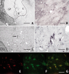
Immunocytochemical staining of TASK-3 channels in cholinergic neurons of the basal nucleus of Meynert a Immunocytochemical staining of the basal forebrain (Bregma: −1.4 mm) with an anti-vesicular acetylcholine transporter antibody (anti-VAChT) identified cholinergic neurons of the basal nucleus of Meynert. The expression of TASK-3 is visualized at the mRNA b and the protein level c, d. Immunocytochemistry with the anti-TASK-3 antibody c of a section adjacent to a revealed strongly TASK-3-positive neurons in this area. At higher magnification d the localization of TASK-3 protein in somata and dendrites (see arrow in c) is appreciated. Immunofluorescence double-staining with anti-VAChT (E, Cy3, red) and anti-TASK-3 (F, Cy2, green) confirmed that TASK-3 is expressed in cholinergic neurons of the basal nucleus of Meynert (G, merge). B, basal nucleus of Meynert; CPu, caudate putamen; GP, globus pallidus; ic, internal capsule. Bar represent 400 μm in a, 100 μm in b, c, and d, and 50 μm in e, g, and f. (Color figure online)

Similar articles

Cited by

References

    1. Aller MI, Veale EL, Linden AM, Sandu C, Schwaninger M, Evans LJ, Korpi ER, Mathie A, Wisden W, Brickley SG (2005) Modifying the subunit composition of TASK channel alters the modulation of a leak conductance in cerebellar granule neurons. J Neurosci 25:11455–11467 - PMC - PubMed
    1. Bayliss DA, Talley EM, Sirois JE, Lei Q (2001) TASK-1 is a highly modulated pH-sensitive ‘leak’ K(+) channel expressed in brainstem respiratory neurons. Respr Physiol 129:159–174 - PubMed
    1. Ben-Shahar O, Obara I, Ary AW, Ma N, Mangiardi MA, Medina RL, Szumlinski KK (2009) Extended daily access to cocaine results in distinct alterations in Homer 1b/c and NMDA receptor subunit expression within the medial prefrontal cortex. Synapse 63:598–609 - PMC - PubMed
    1. Berg AP, Talley EM, Manger JP, Bayliss DA (2004) Motoneurons express heteromeric TWIK-related acid-sensitive K+ (TASK) channels containing TASK-1 (KCNK3) and TASK-3 (KCNK9) subunits. J Neurosci 24:6693–6702 - PMC - PubMed
    1. Bird MK, Reid CA, Chen F, Tan HO, Petrou S, Lawrence AJ (2009) Cocaine-mediated synaptic potentiation is absent in VTA neurons from mGlu5-deficient mice. Int J Neuropsychopharmacol 13:133–141 - PubMed

MeSH terms

LinkOut - more resources