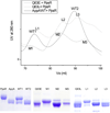
A Q63E Rhodobacter sphaeroides AppA BLUF domain mutant is locked in a pseudo-light-excited signaling state
- PMID: 21082791
- PMCID: PMC3127458
- DOI: 10.1021/bi1002162
A Q63E Rhodobacter sphaeroides AppA BLUF domain mutant is locked in a pseudo-light-excited signaling state
Abstract
The AppA BLUF photoreceptor from Rhodobacter sphaeroides contains a conserved key residue, Gln63, that is thought to undergo a shift in hydrogen-bonding interactions when a bound flavin is light excited. In this study we have characterized two substitution mutants of Gln63 (Q63E, Q63L) in the context of two constructs of the BLUF domain that have differing lengths, AppA1-126 and AppA17-133. Q63L mutations in both constructs exhibit a blue-shifted flavin absorption spectrum as well as a loss of the photocycle. Altered fluorescence emission and fluorescence quenching of the Q63L mutant indicate significant perturbations of hydrogen bonding to the flavin and surrounding amino acids which is confirmed by (1)H-(15)N HSQC NMR spectroscopy. The Q63E substitution mutant is constitutively locked in a lit signaling state as evidenced by a permanent 3 nm red shift of the flavin absorption, quenching of flavin fluorescence emission, analysis of (1)H-(15)N HSQC spectra, and the inability of full-length AppA Q63E to bind to the PpsR repressor. The significance of these findings on the mechanism of light-induced output signaling is discussed.
Figures


References
-
- Masuda S, Hasegawa K, Ishii A, Ono TA. Light-induced structural changes in a putative blue-light receptor with a novel FAD binding fold sensor of blue-light using FAD (BLUF); Slr1694 of synechocystis sp. PCC6803. Biochemistry. 2004;43:5304–5313. - PubMed
-
- Hasegawa K, Masuda S, Ono TA. Structural intermediate in the photocycle of a BLUF (sensor of blue light using FAD) protein Slr1694 in a Cyanobacterium Synechocystis sp. PCC6803. Biochemistry. 2004;43:14979–14986. - PubMed
-
- Kita A, Okajima K, Morimoto Y, Ikeuchi M, Miki K. Structure of a cyanobacterial BLUF protein, Tll0078, containing a novel FAD-binding blue light sensor domain. J Mol Biol. 2005;349:1–9. - PubMed
-
- Rajagopal S, Key JM, Purcell EB, Boerema DJ, Moffat K. Purification and initial characterization of a putative blue light-regulated phosphodiesterase from Escherichia coli. Photochem Photobiol. 2004;80:542–547. - PubMed
Publication types
MeSH terms
Substances
Grants and funding
LinkOut - more resources
Full Text Sources

