

A diffusion tensor imaging study on the white matter skeleton in individuals with sports-related concussion
- PMID: 21083414
- PMCID: PMC3037804
- DOI: 10.1089/neu.2010.1430
A diffusion tensor imaging study on the white matter skeleton in individuals with sports-related concussion
Abstract
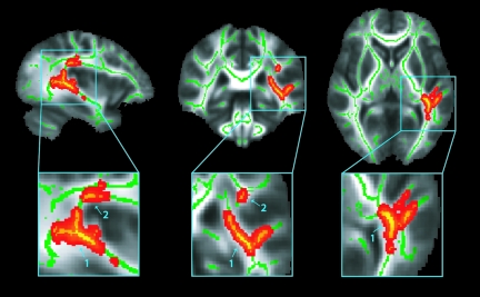
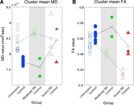
Recognizing and managing the effects of cerebral concussion is very challenging, given the discrete symptomatology. Most individuals with sports-related concussion will not score below 15 on the Glasgow Coma Scale, but will present with rapid onset of short-lived neurological impairment, demonstrating no structural changes on traditional magnetic resonance imaging (MRI) and computed tomography (CT) scans. The return-to-play decision is one of the most difficult responsibilities facing the physician, and so far this decision has been primarily based on neurological examination, symptom checklists, and neuropsychological (NP) testing. Diffusion tensor imaging (DTI) may be a more objective tool to assess the severity and recovery of function after concussion. We assessed white matter (WM) fiber tract integrity in varsity level college athletes with sports-related concussion without loss of consciousness, who experienced protracted symptoms for at least 1 month after injury. Evaluation of fractional anisotropy (FA) and mean diffusivity (MD) of the WM skeleton using tract-based spatial statistics (TBSS) revealed a large cluster of significantly increased MD for concussed subjects in several WM fiber tracts in the left hemisphere, including parts of the inferior/superior longitudinal and fronto-occipital fasciculi, the retrolenticular part of the internal capsule, and posterior thalamic and acoustic radiations. Qualitative comparison of average FA and MD suggests that with increasing level of injury severity (ranging from sports-related concussion to severe traumatic brain injury), MD might be more sensitive at detecting mild injury, whereas FA captures more severe injuries. In conclusion, the TBSS analysis used to evaluate diffuse axonal injury of the WM skeleton seems sensitive enough to detect structural changes in sports-related concussion.
Figures
References
-
- Acosta-Cabronero J. Williams G.B. Pengas G. Nestor P.J. Absolute diffusivities define the landscape of white matter degeneration in Alzheimer's disease. Brain. 2010;133:529–539. - PubMed
-
- Anderson B. Southern B.D. Powers R.E. Anatomic asymmetries of the posterior superior temporal lobes: A post-mortem study. Neuropsychiatry Neuropsychol. Behav. Neurol. 1999;12:247–254. - PubMed
-
- Andersson J.L.R. Jenkinson M. Smith S. Non-linear optimization. FMRIB technical report TR07JA1. 2007a. www.fmrib.ox.ac.uk/analyis/techrep. www.fmrib.ox.ac.uk/analyis/techrep from.
-
- Andersson J.L.R. Jenkinson M. Smith S. Non-linear registration, aka Spatial normalization. FMRIB technical report TR07JA2. 2007b. www.fmrib.ox.ac.uk/analysis/techrep. www.fmrib.ox.ac.uk/analysis/techrep from.
Publication types
MeSH terms
Grants and funding
LinkOut - more resources
Full Text Sources
Other Literature Sources
Medical
Miscellaneous
