. 2011 Feb;28(2):299-309.
doi: 10.1089/neu.2010.1594. Epub 2011 Jan 9.

Activity-dependent increase in neurotrophic factors is associated with an enhanced modulation of spinal reflexes after spinal cord injury

Affiliations

Activity-dependent increase in neurotrophic factors is associated with an enhanced modulation of spinal reflexes after spinal cord injury

Marie-Pascale Côté et al. J Neurotrauma. 2011 Feb.

Abstract

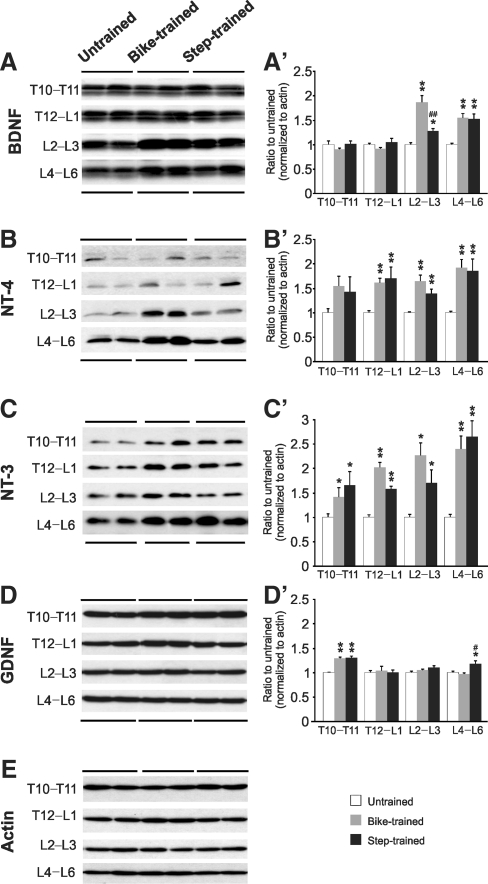
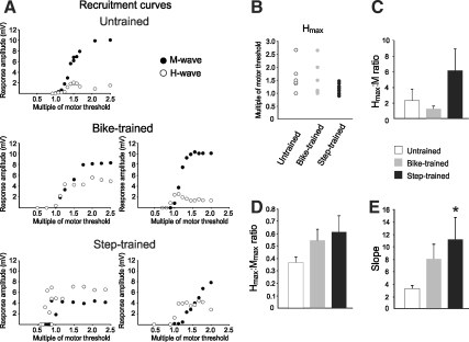
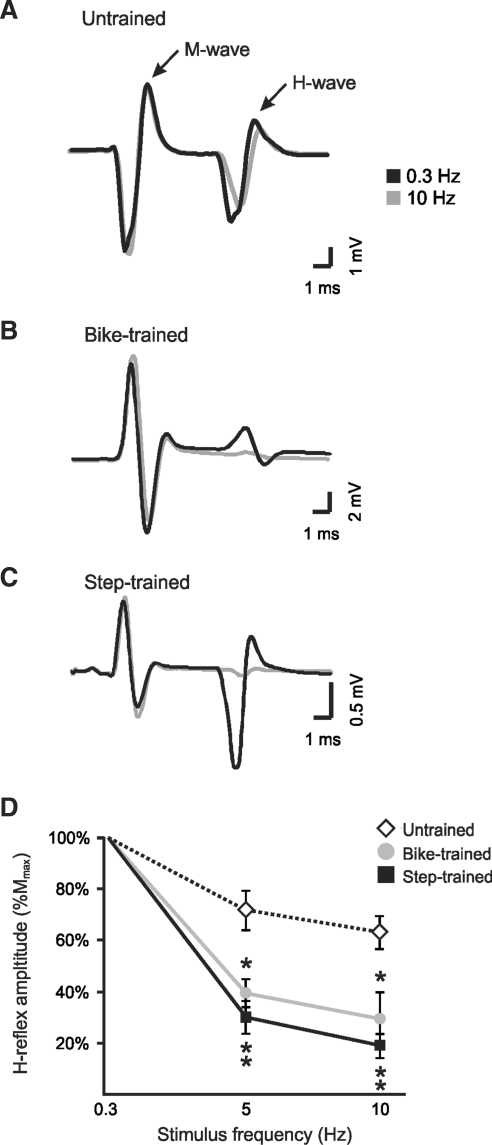
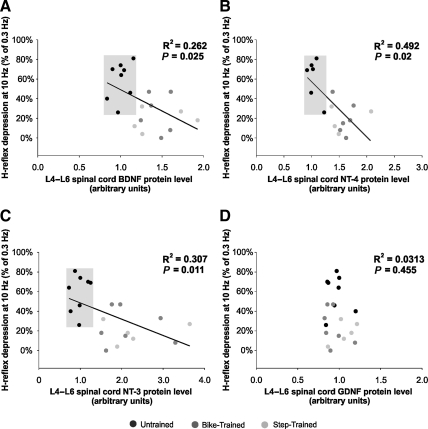
Activity-based therapies such as passive bicycling and step-training on a treadmill contribute to motor recovery after spinal cord injury (SCI), leading to a greater number of steps performed, improved gait kinematics, recovery of phase-dependent modulation of spinal reflexes, and prevention of decrease in muscle mass. Both tasks consist of alternating movements that rhythmically stretch and shorten hindlimb muscles. However, the paralyzed hindlimbs are passively moved by a motorized apparatus during bike-training, whereas locomotor movements during step-training are generated by spinal networks triggered by afferent feedback. Our objective was to compare the task-dependent effect of bike- and step-training after SCI on physiological measures of spinal cord plasticity in relation to changes in levels of neurotrophic factors. Thirty adult female Sprague-Dawley rats underwent complete spinal transection at a low thoracic level (T12). The rats were assigned to one of three groups: bike-training, step-training, or no training. The exercise regimen consisted of 15 min/d, 5 days/week, for 4 weeks, beginning 5 days after SCI. During a terminal experiment, H-reflexes were recorded from interosseus foot muscles following stimulation of the tibial nerve at 0.3, 5, or 10 Hz. The animals were sacrificed and the spinal cords were harvested for Western blot analysis of the expression of neurotrophic factors in the lumbar spinal cord. We provide evidence that bike- and step-training significantly increase the levels of brain-derived neurotrophic factor (BDNF), neurotrophin-3 (NT-3), and NT-4 in the lumbar enlargement of SCI rats, whereas only step-training increased glial cell-derived neurotrophic factor (GDNF) levels. An increase in neurotrophic factor protein levels that positively correlated with the recovery of H-reflex frequency-dependent depression suggests a role for neurotrophic factors in reflex normalization.


Figures

FIG. 1.
FIG. 1.
Western blots showing the expression of brain-derived neurotrophic factor (BDNF) (A), neurotrophin-4 (NT-4) (B), NT-3 (C), and glial cell-derived neurotrophic factor (GDNF) (D) rostral to the injury site (T10–T11), at the lesion site (T12–L1), and caudal to the injury site (L2–L3 and L4–L6), in untrained, bike-trained, and step-trained animals (n = 8 per group). Overall, bike- and step-training increased the expression of BDNF (A’), NT-3 (B’), NT-4 (C’), and GDNF (D’). Also, step-training increased GDNF protein levels in the lumbar enlargement more than bike-training (D’). The blots were reprobed with anti-actin to provide an indication of total protein loaded per lane (E). BDNF, NT-3, NT-4, and GDNF proteins were detected and quantified by Western blots using actin as a standard control (*p < 0.05 versus untrained; **p < 0.001 versus untrained; #p < 0.05 versus bike-trained; ##p < 0.001 versus bike-trained).
FIG. 2.
FIG. 2.
(A) Representative recruitment curves for individual animals in the untrained, bike-trained, and step-trained groups. The untrained animals display Hmax ∼1.5 MT, and Mmax ∼2 MT. Bike- and step-training shifted the Hmax value toward 1 MT. (B) Hmax occurrence expressed as a multiple of MT. (C) The Hmax:M ratio tends to increase with step-training, suggesting that Hmax occurs when M is of smaller amplitude than in untrained animals. (D) The Hmax:Mmax ratio tends to increase with exercise. (E) The slope of the recruitment curve is significantly steeper in step-trained animals than in untrained animals (MT, motor threshold; Mmax, maximal response amplitude for the M-wave; Hmax, maximal response amplitude for the H-wave).
FIG. 3.
FIG. 3.
H-reflex frequency-dependent depression is recovered after 30 days of step-training and bike-training in spinal cord injury (SCI) animals. H-reflexes were evoked by the stimulation of the tibial nerve and recorded from the interosseus muscles. Representative averages of H-reflex recordings following a train of stimulation at 0.3 Hz (black), and 10 Hz (gray), in untrained (A), bike-trained (B), and step-trained (C) animals. There was a modest decrease in the average amplitude of the H-reflex at 10 Hz compared to 0.3 Hz in untrained animals, whereas this decrease was considerable in bike-trained and step-trained animals. Overall, the animals in the exercised groups showed a marked reduction of the H-reflex amplitude as the stimulation frequency increased (i.e., increased depression of the reflex), compared to the untrained group (D). There was a significant decrease in H-reflex amplitude in bike-trained and step-trained animals compared to untrained animals at both 5 Hz and 10 Hz (D; *p < 0.05; **p < 0.001).
FIG. 4.
FIG. 4.
Linear regression analysis between H-reflex depression and brain-derived neurotrophic factor (BDNF) (A), neurotrophin-4 (NT-4) (B), NT-3 (C), and glial cell-derived neurotrophic factor (GDNF) (D) protein levels in the L4–L6 spinal segments. There was a significant correlation between BDNF, NT-3, and NT-4 levels, and the depth of the H-reflex depression at 10 Hz (expressed as a percentage of 0.3 Hz). This correlation was not observed with GDNF. Untrained animals are segregated in the upper left quadrant, and are linearly separable from the exercised groups (gray boxes), for BDNF, NT-3, and NT-4, expressed as a function of H-reflex depression, but GDNF is not.

Similar articles

Cited by

References

    1. Beaumont E. Houlé J.D. Peterson C.A. Gardiner P.F. Passive exercise and fetal spinal cord transplant both help to restore motoneuronal properties after spinal cord transection in rats. Muscle Nerve. 2004;29:234–242. - PubMed
    1. Beaumont E. Kaloustian S. Rousseau G. Cormery B. Training improves the electrophysiological properties of lumbar neurons and locomotion after thoracic spinal cord injury in rats. Neurosci. Res. 2008;62:147–154. - PubMed
    1. Bennett D.J. Li Y. Harvey P.J. Gorassini M. Evidence for plateau potentials in tail motoneurons of awake chronic spinal rats with spasticity. J. Neurophysiol. 2001;86:1972–1982. - PubMed
    1. Boorman G. Becker W.J. Morrice B.L. Lee R.G. Modulation of the soleus H-reflex during pedalling in normal humans and in patients with spinal spasticity. J. Neurol. Neurosurg. Psychiatry. 1992;55:1150–1156. - PMC - PubMed
    1. Boulenguez P. Liabeuf S. Bos R. Bras H. Jean-Xavier C. Brocard C. Stil A. Darbon P. Cattaert D. Delpire E. Marsala M. Vinay L. Down-regulation of the potassium-chloride cotransporter KCC2 contributes to spasticity after spinal cord injury. Nat. Med. 2010;16:302–307. - PubMed

Publication types

Substances