. 2010 Oct;15(5):403-15.
doi: 10.1111/j.1523-5378.2010.00770.x.

Morphologic, genetic, and biochemical characterization of Helicobacter magdeburgensis, a novel species isolated from the intestine of laboratory mice

Affiliations

Morphologic, genetic, and biochemical characterization of Helicobacter magdeburgensis, a novel species isolated from the intestine of laboratory mice

Francisco Rivas Traverso et al. Helicobacter. 2010 Oct.

Abstract

Background: The presence of enterohepatic Helicobacter species (EHS) is commonly noted in mouse colonies. These infections often remain unrecognized but can cause severe health complications or more subtle host immune perturbations and therefore can confound the results of animal experiments. The aim of this study was to isolate and characterize a putative novel EHS that has previously been detected by PCR screening of specific-pathogen-free mice.

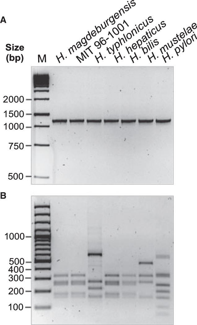
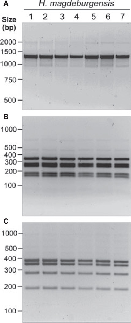
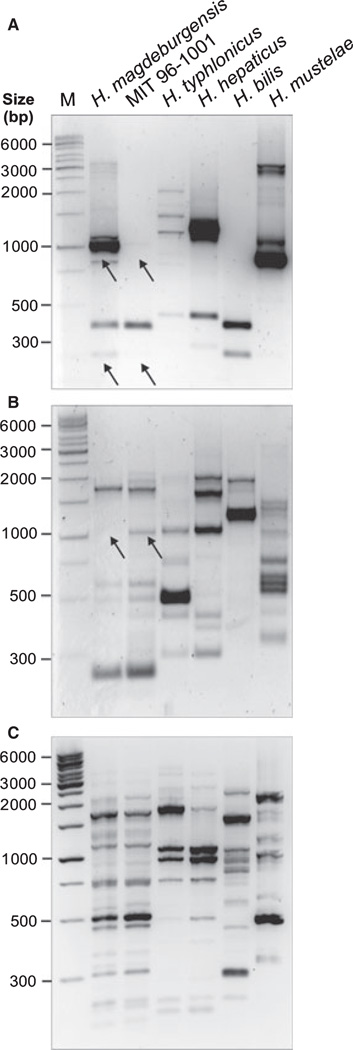
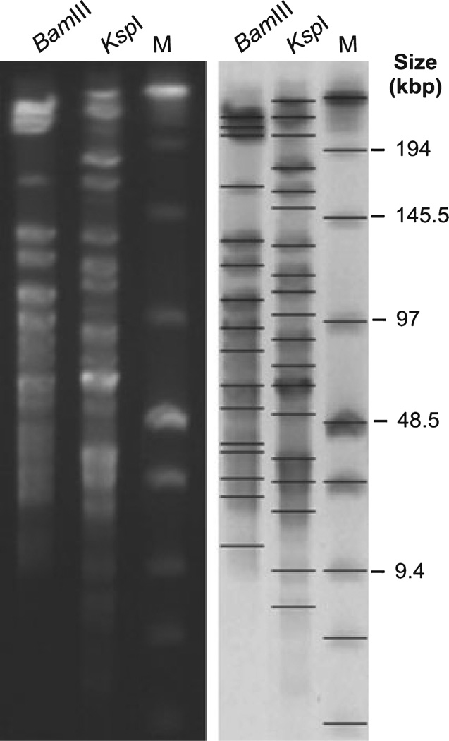
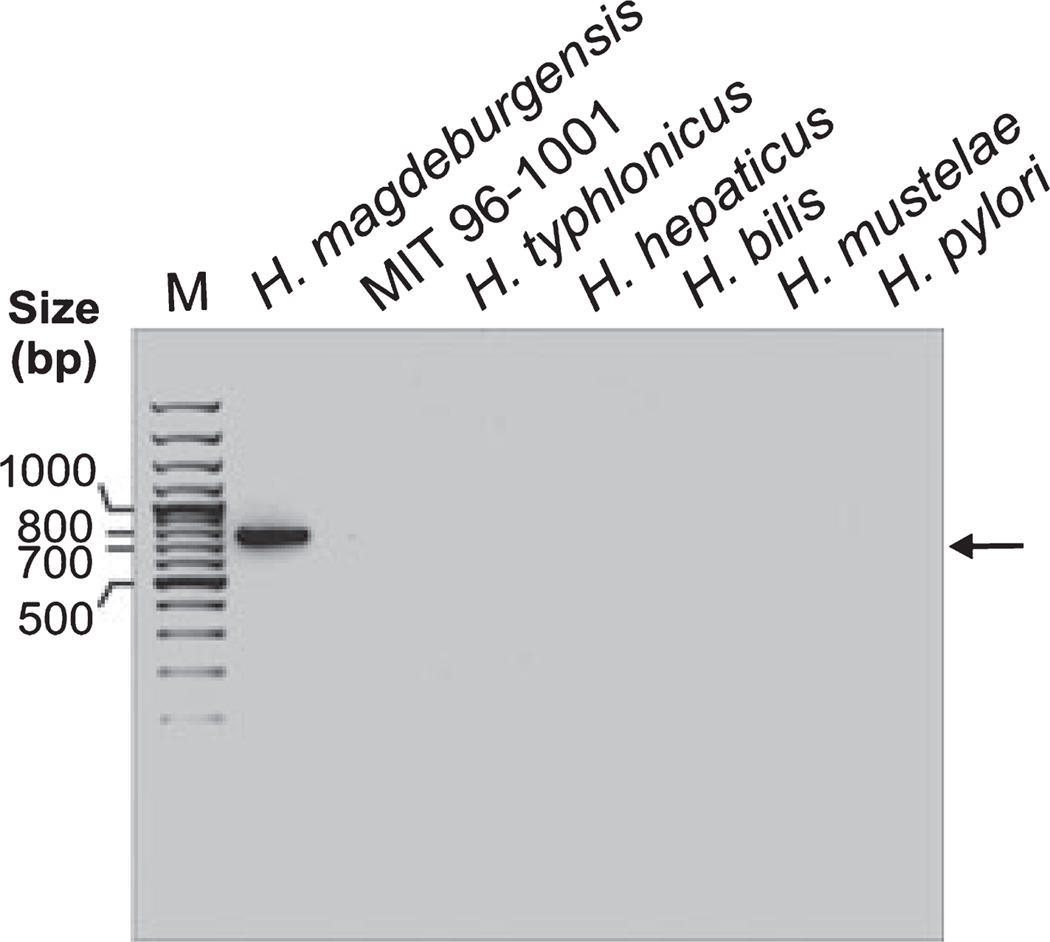
Materials and methods: Biochemical analysis of enzyme activities (API campy), morphologic investigation (Gram-staining and electron microscopy) and genetic analyses (16SrRNA and 23SrRNA analyses, DNA fingerprinting, restriction fragment polymorphisms, and pulsed-field gel electrophoresis) were used to characterize isolated EHS. Genomic DNA fragments were sequenced to develop a species-specific PCR detection assay.

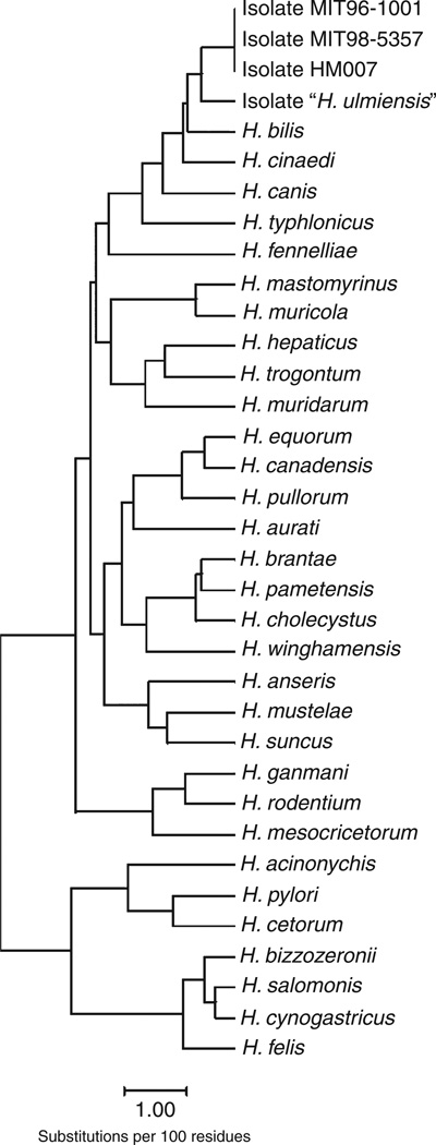
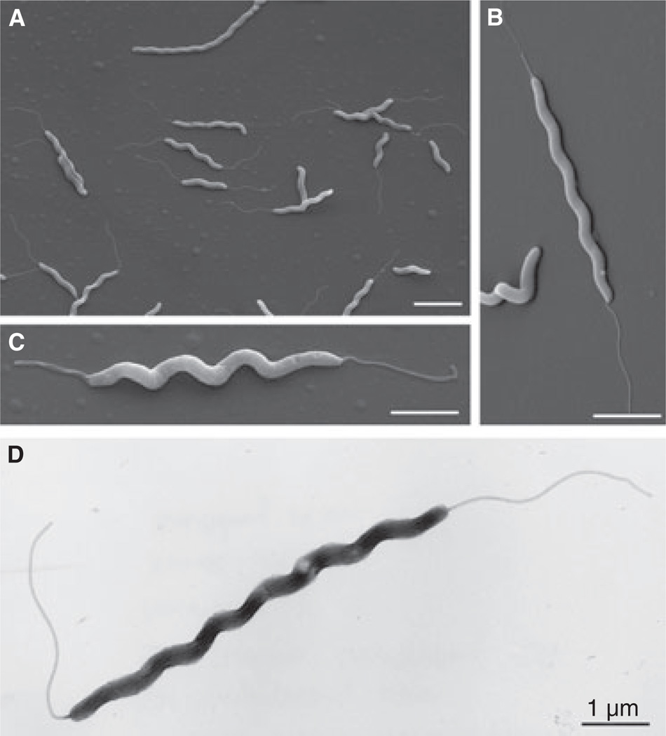
Results: Scanning electron microscopy revealed the presence of spiral-shaped EHS, which varied in length (2.5-6 μm) and contained single monopolar or single bipolar sheathed flagella. The bacteria were grown under anaerobic conditions, preferably on agar plates containing serum or blood. The 16SrRNA, genetic, and biochemical analyses indicated the identification of a novel EHS species, named Helicobacter magdeburgensis. We also examined the genome content using pulsed-field gel electrophoresis. Based on the pattern produced by two restriction enzymes, BamIII and KspI, the genome size was determined to be about 1.7-1.8 Mbp.

Conclusion: We isolated and characterized a novel EHS species, H. magdeburgensis, morphologically, biochemically, and genetically. These results are important for future studies on the prevalence and pathophysiologic relevance of such infections. Our PCR assay can be used to detect and discriminate H. magdeburgensis from other Helicobacter species.


Figures

Figure 1
Figure 1
Investigation of 16S rRNA genes of different Helicobacter species by PCR and RFLP analyses. (A) DNA isolated from bacteria belonging to the genus Helicobacter (H. magdeburgensis, H. typhlonicus, H. hepaticus, H. bills, H. mustelae, H. pylori, and the MIT strain 96-1001) was applied for conventional PCR of the 16S rRNA gene. A conserved 1.2-kb DNA fragment in the genus Helicobacter was amplified [28]. (B) To confirm the specificity of these fragments, all PCR products were then digested with the restriction endonuclease AluI, which gives raise to a specific band pattern as described [25].
Figure 2
Figure 2
Analysis of 16S rRNA of different Helicobacter magdeburgensis isolates by PCR and RFLPs. (A) DNA isolated from seven individual clones belonging to H. magdeburgensis was investigated by conventional PCR of the 16S rRNA gene. A conserved 1.2-kb DNA fragment in the genus Helicobacter was amplified [28]. To confirm the specificity of these fragments, all PCR products were then digested with the restriction endonuclease AluI (B) or HhaI (C) gives raise to a specific band pattern as described [25] and was identical among all investigated clones.
Figure 3
Figure 3
Phylogenetic tree of 16S rRNA sequences of different Helicobacter species including H. magdeburgensis.
Figure 4
Figure 4
Morphologic analyses of novel Helicobacter species by electron microscopy. (A–C) Scanning electron microscopy revealed spiral-shaped bacteria that were about 0.18–0.22 µm in diameter and varied in length from about 2.5–6 µm. The majority of bacteria contained single monopolar or single bipolar flagella. Representative pictures are shown from three preparations. (D) Investigation of the Helicobacter species by another method (negative staining) revealed similar results with respect to size and morphology. Each bar corresponds to 1 µm.
Figure 5
Figure 5
PCR-based randomly amplified polymorphic DNA (RAPD) fingerprinting of Helicobacter species. To investigate the genetic relatedness between H. magdeburgensis and the closest known relative (MIT 96-1001) and other strains, we performed RAPD analysis as described [37]. (A–C) This method uses a set of single primers (D14307, D9355 or D8635), which arbitrarily anneal and amplify genomic DNA resulting in strain-specific fingerprinting patterns [39]. Typical RAPD fingerprinting profiles with each of the three primers are shown. Arrows indicate some bands either present or missing in H. magdeburgensis and MIT 96-1001, respectively.
Figure 6
Figure 6
Pulsed-field gel electrophoresis (PFGE) analysis of Helicobacter magdeburgensis. Chromosomal DNA was digested with the restriction endonucleases BamIII and KspI, respectively. Low Range PFGE Marker was used as the DNA size marker (M). BioNumerics software was used to identify bands and to determine band sizes. The values from the genome calculations are summarized in Table 2.
Figure 7
Figure 7
Development of a H. magdeburgensis-specific PCR detection assay. We designed specific PCR primers as described in the Materials and Methods section. Using these primers, we developed a PCR assay giving rise to a single H. magdeburgensis-specific 750-bp DNA fragment, which is clearly absent in all other Helicobacter isolates tested, even after 35 PCR cycles, including the MIT strain 96-1001, H. typhlonicus, H. hepaticus, H. bills, H. mustelae, and H. pylori.

Similar articles

Cited by

References

    1. Dewhirst FE, Fox JG, On SL. Recommended minimal standards for describing new species of the genus Helicobacter. Int J Syst Evol Microbiol. 2000;50:2231–2237. - PubMed
    1. Marshall BJ, Warren JR. Unidentified curved bacilli in the stomach of patients with gastritis and peptic ulceration. Lancet. 1984;1:1311–1315. - PubMed
    1. Marshall BJ, Royce H, Annear DI, et al. Original isolation of Campylobacter pyloridis from human gastric mucosa. Microbios. 1984;25:83–88.
    1. Romaniuk PJ, Zoltowska B, Trust TJ, et al. Campylobacter pylori, the spiral bacterium associated with human gastritis, is not a true Campylobacter sp. J Bacterial. 1987;169:2137–2141. - PMC - PubMed
    1. Paster BJ, Dewhirst FE. Phylogeny of Campylobacter, wolinellas, Bacteroides gracilis, and Bacteroides ureolyticus by 16S ribosomal ribonucleic acid sequencing. Int J Syst Bacteriol. 1988;38:56–62.

Publication types

MeSH terms

Associated data

LinkOut - more resources