

Segmental cholangitis impairs hepatic regeneration capacity after partial hepatectomy in rats
- PMID: 21083791
- PMCID: PMC3003476
- DOI: 10.1111/j.1477-2574.2010.00229.x
Segmental cholangitis impairs hepatic regeneration capacity after partial hepatectomy in rats
Abstract
Background: Patients with hilar cholangiocarcinoma or hepatolithiasis often develop segmental cholangitis (SC), but it is unclear whether hepatectomy for patients with SC can be performed safely.
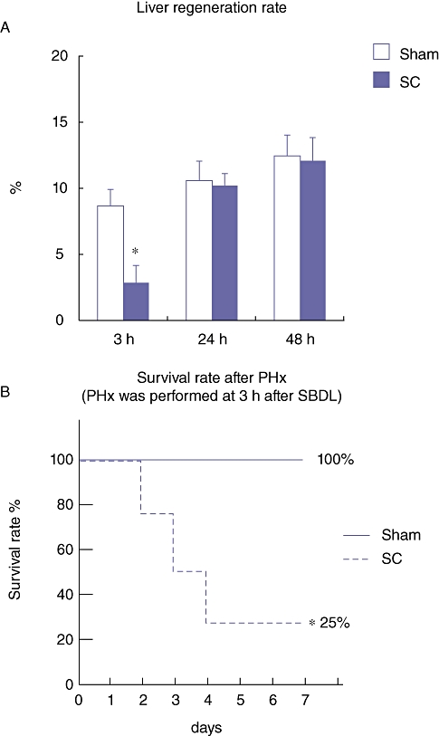
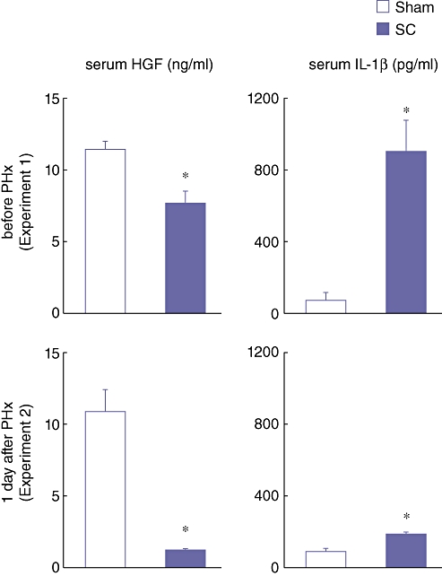
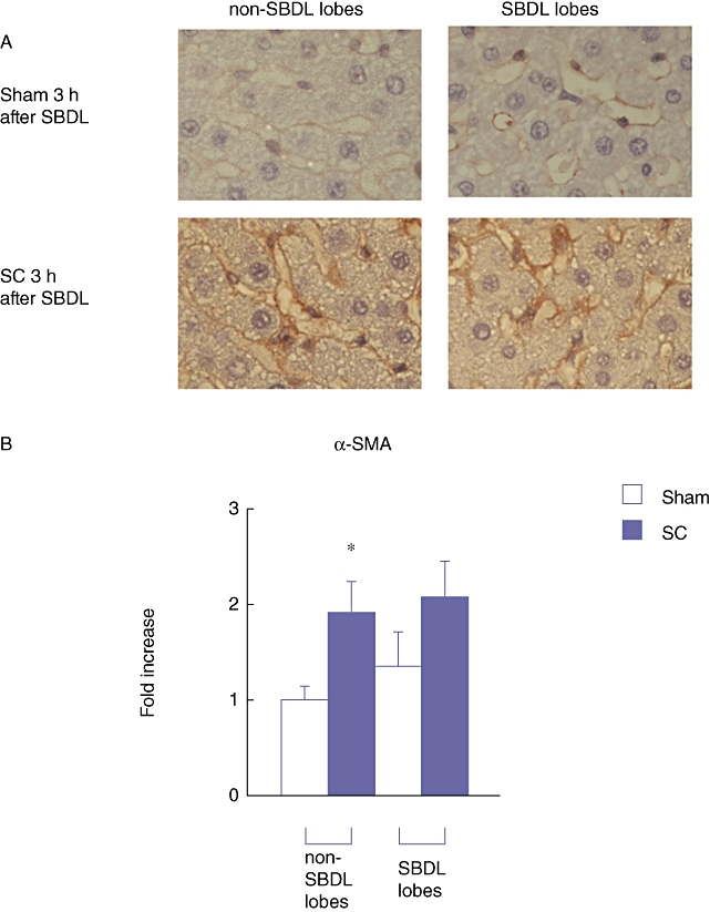
Methods: Rats were subjected to segmental bile duct ligation (SBDL) with LPS (SC group) or a saline (Sham group) infusion into the bile duct of the ligated lobes. The rats were sacrificed at 3, 24 and 48 h after the SBDL. For another experiment, the rats were subjected to partial hepatectomy (PHx) for the ligated lobes. Hepatic regeneration rates and the expression of regeneration-associated genes were evaluated.
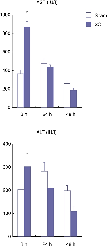
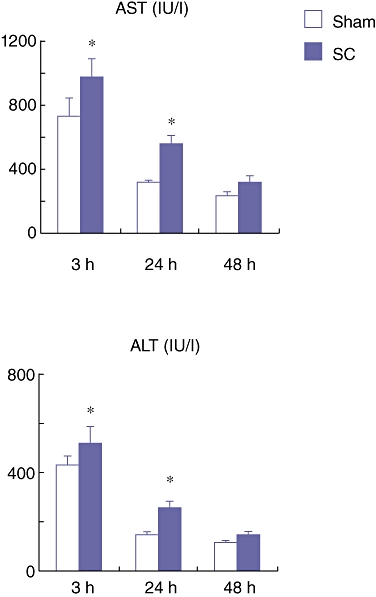
Results: In the SC group, severe parenchymal damage was observed in the acute phase (3 h). Altered gene expression in the liver in response to biliary infection occurred not only in the infected lobes but also in the non-infected lobes. In the rats of the SC group, both the hepatic regeneration rate and serum HGF levels were significantly lower than in the Sham group.
Conclusion: These results clearly demonstrate that SC impairs the regeneration capacity of the contralateral remnant liver. Therefore, hepatectomy should be avoided for patients with SC even if it occurs in the part of the liver to be resected.
Figures


Similar articles
-
Is there any effect of renal failure on the hepatic regeneration capacity following partial hepatectomy in rats?Biochem Biophys Res Commun. 2007 Jan 12;352(2):311-6. doi: 10.1016/j.bbrc.2006.11.011. Epub 2006 Nov 13. Biochem Biophys Res Commun. 2007. PMID: 17126297
-
Liver regeneration following experimental major hepatectomy with choledochojejunostomy.Br J Surg. 2015 Oct;102(11):1410-7. doi: 10.1002/bjs.9908. Epub 2015 Aug 27. Br J Surg. 2015. PMID: 26312457
-
15-deoxy-delta 12,14-prostaglandin J2 prevents inflammatory response and endothelial cell damage in rats with acute obstructive cholangitis.Am J Physiol Gastrointest Liver Physiol. 2010 Mar;298(3):G410-8. doi: 10.1152/ajpgi.00233.2009. Epub 2010 Jan 7. Am J Physiol Gastrointest Liver Physiol. 2010. PMID: 20056897
-
A modified animal model of hepatic regeneration induced by hilar bile duct ligation.Sci Rep. 2021 Oct 12;11(1):20201. doi: 10.1038/s41598-021-99758-z. Sci Rep. 2021. PMID: 34642435 Free PMC article.
-
Mechanism of liver regeneration after liver resection and portal vein embolization (ligation) is different?J Hepatobiliary Pancreat Surg. 2009;16(3):292-9. doi: 10.1007/s00534-009-0058-x. Epub 2009 Mar 31. J Hepatobiliary Pancreat Surg. 2009. PMID: 19333540 Review.
Cited by
-
Preoperative Cholangitis and Future Liver Remnant Volume Determine the Risk of Liver Failure in Patients Undergoing Resection for Hilar Cholangiocarcinoma.J Am Coll Surg. 2016 Jul;223(1):87-97. doi: 10.1016/j.jamcollsurg.2016.01.060. Epub 2016 Feb 13. J Am Coll Surg. 2016. PMID: 27049784 Free PMC article.
-
Liver Regeneration after Hepatectomy and Partial Liver Transplantation.Int J Mol Sci. 2020 Nov 9;21(21):8414. doi: 10.3390/ijms21218414. Int J Mol Sci. 2020. PMID: 33182515 Free PMC article. Review.
-
External biliary drainage following major liver resection for perihilar cholangiocarcinoma: impact on development of liver failure and biliary leakage.HPB (Oxford). 2016 Apr;18(4):348-53. doi: 10.1016/j.hpb.2015.11.007. Epub 2016 Feb 18. HPB (Oxford). 2016. PMID: 27037204 Free PMC article.
-
Update on Liver Failure Following Hepatic Resection: Strategies for Prediction and Avoidance of Post-operative Liver Insufficiency.J Clin Transl Hepatol. 2018 Mar 28;6(1):97-104. doi: 10.14218/JCTH.2017.00060. Epub 2017 Nov 30. J Clin Transl Hepatol. 2018. PMID: 29577036 Free PMC article. Review.
References
-
- Chen DW, Tung-Ping Poon R, Liu CL, Fan ST, Wong J. Immediate and long-term outcomes of hepatectomy for hepatolithiasis. Surgery. 2004;135:386–393. - PubMed
-
- Nagino M, Nimura Y, Hayakawa N, Kamiya J, Kondo S, Sasaki R, et al. Logistic regression and discriminant analyses of hepatic failure after liver resection for carcinoma of the biliary tract. World J Surg. 1993;17:250–255. - PubMed
-
- Kanai M, Nimura Y, Kamiya J, Kondo S, Nagino M, Miyachi M, et al. Preoperative intrahepatic segmental cholangitis in patients with advanced carcinoma involving the hepatic hilus. Surgery. 1996;119:498–504. - PubMed
MeSH terms
Substances
LinkOut - more resources
Full Text Sources
