

Contribution of constitutively proliferating precursor cell subtypes to dentate neurogenesis after cortical infarcts
- PMID: 21083887
- PMCID: PMC2993721
- DOI: 10.1186/1471-2202-11-146
Contribution of constitutively proliferating precursor cell subtypes to dentate neurogenesis after cortical infarcts
Abstract
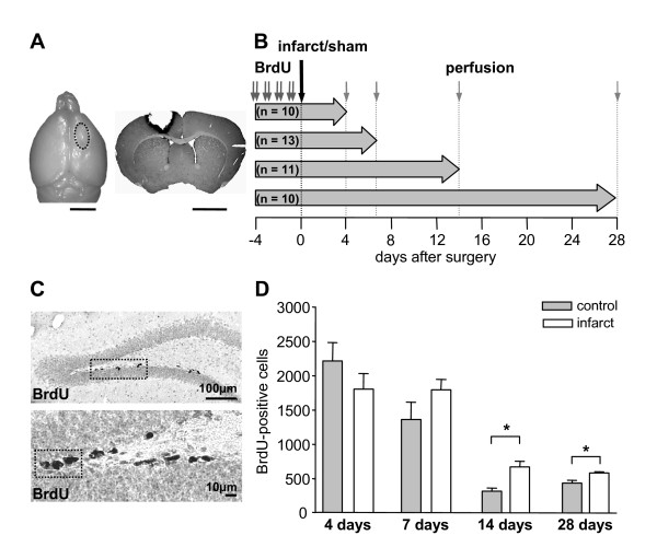
Background: It is well known that focal ischemia increases neurogenesis in the adult dentate gyrus of the hippocampal formation but the cellular mechanisms underlying this proliferative response are only poorly understood. We here investigated whether precursor cells which constitutively proliferate before the ischemic infarct contribute to post-ischemic neurogenesis. To this purpose, transgenic mice expressing green fluorescent protein (GFP) under the control of the nestin promoter received repetitive injections of the proliferation marker bromodeoxyuridine (BrdU) prior to induction of cortical infarcts. We then immunocytochemically analyzed the fate of these BrdU-positive precursor cell subtypes from day 4 to day 28 after the lesion.
Results: Quantification of BrdU-expressing precursor cell populations revealed no alteration in number of radial glia-like type 1 cells but a sequential increase of later precursor cell subtypes in lesioned animals (type 2a cells at day 7, type 3 cells/immature neurons at day 14). These alterations result in an enhanced survival of mature neurons 4 weeks postinfarct.
Conclusions: Focal cortical infarcts recruit dentate precursor cells generated already before the infarct and significantly contribute to an enhanced neurogenesis. Our findings thereby increase our understanding of the complex cellular mechanisms of postlesional neurogenesis.
Figures


Similar articles
-
Differential stroke-induced proliferative response of distinct precursor cell subpopulations in the young and aged dentate gyrus.Neuroscience. 2010 Sep 1;169(3):1279-86. doi: 10.1016/j.neuroscience.2010.05.035. Epub 2010 May 24. Neuroscience. 2010. PMID: 20570606
-
Neurogenesis in the adult dentate gyrus after cortical infarcts: effects of infarct location, N-methyl-D-aspartate receptor blockade and anti-inflammatory treatment.Neuroscience. 2005;135(3):723-35. doi: 10.1016/j.neuroscience.2005.06.082. Epub 2005 Sep 8. Neuroscience. 2005. PMID: 16154293
-
Temporal profile of neurogenesis in the subventricular zone, dentate gyrus and cerebral cortex following transient focal cerebral ischemia.Neurol Res. 2009 Nov;31(9):969-76. doi: 10.1179/174313209X383312. Epub 2009 Jan 9. Neurol Res. 2009. PMID: 19138475
-
Adult neurogenesis and the plasticity of the dentate gyrus network.Eur J Neurosci. 2011 Mar;33(6):1055-61. doi: 10.1111/j.1460-9568.2011.07603.x. Eur J Neurosci. 2011. PMID: 21395848 Review.
-
Review: adult neurogenesis contributes to hippocampal plasticity.Cell Tissue Res. 2018 Sep;373(3):693-709. doi: 10.1007/s00441-017-2735-4. Epub 2017 Nov 29. Cell Tissue Res. 2018. PMID: 29185071 Review.
Cited by
-
Post-Stroke Environmental Enrichment Improves Neurogenesis and Cognitive Function and Reduces the Generation of Aberrant Neurons in the Mouse Hippocampus.Cells. 2023 Feb 17;12(4):652. doi: 10.3390/cells12040652. Cells. 2023. PMID: 36831319 Free PMC article.
-
Adult Neurogenesis and Stroke: A Tale of Two Neurogenic Niches.Front Neurosci. 2021 Aug 10;15:700297. doi: 10.3389/fnins.2021.700297. eCollection 2021. Front Neurosci. 2021. PMID: 34447293 Free PMC article. Review.
-
Early Stroke Induces Long-Term Impairment of Adult Neurogenesis Accompanied by Hippocampal-Mediated Cognitive Decline.Cells. 2019 Dec 17;8(12):1654. doi: 10.3390/cells8121654. Cells. 2019. PMID: 31861141 Free PMC article.
-
Enhanced neurogenesis after ischemic stroke: The interplay between endogenous and exogenous stem cells.Neural Regen Res. 2026 Jan 1;21(1):212-223. doi: 10.4103/NRR.NRR-D-24-00879. Epub 2025 Jan 13. Neural Regen Res. 2026. PMID: 39820432 Free PMC article.
-
Post-stroke Neurogenesis: Friend or Foe?Front Cell Dev Biol. 2021 Mar 23;9:657846. doi: 10.3389/fcell.2021.657846. eCollection 2021. Front Cell Dev Biol. 2021. PMID: 33834025 Free PMC article. Review.
References
Publication types
MeSH terms
LinkOut - more resources
Full Text Sources
Medical
Molecular Biology Databases
