Review
. 2010 Nov 17:7:96.
doi: 10.1186/1742-4690-7-96.

Revisiting HIV-1 uncoating

Affiliations
Review

Revisiting HIV-1 uncoating

Nathalie Arhel. Retrovirology. .

Abstract

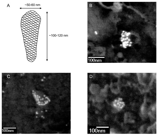
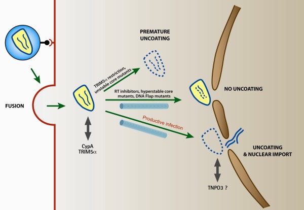
HIV uncoating is defined as the loss of viral capsid that occurs within the cytoplasm of infected cells before entry of the viral genome into the nucleus. It is an obligatory step of HIV-1 early infection and accompanies the transition between reverse transcription complexes (RTCs), in which reverse transcription occurs, and pre-integration complexes (PICs), which are competent to integrate into the host genome. The study of the nature and timing of HIV-1 uncoating has been paved with difficulties, particularly as a result of the vulnerability of the capsid assembly to experimental manipulation. Nevertheless, recent studies of capsid structure, retroviral restriction and mechanisms of nuclear import, as well as the recent expansion of technical advances in genome-wide studies and cell imagery approaches, have substantially changed our understanding of HIV uncoating. Although early work suggested that uncoating occurs immediately following viral entry in the cell, thus attributing a trivial role for the capsid in infected cells, recent data suggest that uncoating occurs several hours later and that capsid has an all-important role in the cell that it infects: for transport towards the nucleus, reverse transcription and nuclear import. Knowing that uncoating occurs at a later stage suggests that the viral capsid interacts extensively with the cytoskeleton and other cytoplasmic components during its transport to the nucleus, which leads to a considerable reassessment of our efforts to identify potential therapeutic targets for HIV therapy. This review discusses our current understanding of HIV uncoating, the functional interplay between infectivity and timely uncoating, as well as exposing the appropriate methods to study uncoating and addressing the many questions that remain unanswered.


Figures

Figure 1
Figure 1
Scanning electron microscopy imaging of HIV-1 capsids in the cytoplasm and at the nuclear membrane of infected cells. (A) Schematic representation of the mature HIV-1 capsid shell. The HIV-1 capsid is an assembly of approximately 1,500 CA monomers arranged into a hexagonal array of hexamers. Dimensions are derived from microscopy observations of mature virions or isolated cores. (B-D) Images show the backscattered gold signal corresponding to specific labelling with a mouse monoclonal anti-p24 antibody (183-H12-5C AIDS Reagent Program) followed with goat anti-mouse IgG H&L conjugated 10 nm gold (British Biocell International) in HIV-1 infected P4-CCR5 cells. HIV-1 capsids are typically conical- or cylindrical-shaped, ca 100-150 nm long, and heavily labelled with 10-30 immunogold particles. The bulk of antibodies likely induces some distortions in size and shape of capsids. In panels B and D, capsids are located at the nuclear membrane: nuclear pore complexes appear as bright rings with dark lumen.
Figure 2
Figure 2
Schematic representation of reverse transcription in lentiviruses and other orthoretroviruses (such as MLV). The conversion of the single-strand RNA genome (represented as a black line) into double stranded DNA genome (at the bottom of the diagram) is the hallmark of retroviruses. Reverse transcription is initiated by the synthesis of minus-strand DNA (in green) at the PBS site (Primer Binding Site) at the 5' end of the RNA genome. The minus-strand strong-stop DNA thus synthesised is then transferred to the 3' end of the genome through complementarity with the R (Repeated) region of the LTR region (Long-Terminal Region) thus allowing synthesis of the minus-strand DNA to be completed. Minus strand DNA synthesis is accompanied by progressive degradation of the RNA matrix by the RNase H activity of reverse transcriptase. Two RNA sequences resist RNase degradation because they contain a unique PPT sequence and these serve as initiation sites for the plus-strand DNA. In all retroviruses, plus-strand DNA synthesis (in red) is initiated at the 3'PPT. In the case of lentiviruses, initiation also takes place at the cPPT. After a second strand transfer, plus-strand DNA synthesis proceeds to generate double-stranded DNA. In the case of lentiviruses, plus-strand initiation in two distinct sites leads to a displacement of the downstream strand over ca 100 nucleotides, terminating at the CTS and thus generating a discrete strand displacement called the central DNA Flap.
Figure 3
Figure 3
Schematic representation of the fates of viral capsids in the cytoplasm of newly infected cells. After entry into the cytoplasm, HIV-1 capsids that are on a path of productive infection remain intact and are transported towards the nucleus along the cytoskeleton. They uncoat at the nuclear membrane upon completion of reverse transcription. Premature uncoating, in the case of TRIM5α restriction or of unstable capsid mutants, leads to abortive infection. Similarly, compromised uncoating, in the case of incomplete reverse transcription or of hyperstable capsid mutants, also leads to a dead-end infection event.

References

    1. Höglund S, Ofverstedt LG, Nilsson A, Lundquist P, Gelderblom H, Ozel M, Skoglund U. Spatial visualization of the maturing HIV-1 core and its linkage to the envelope. AIDS Res Hum Retroviruses. 1992;8:1–7. doi: 10.1089/aid.1992.8.1. - DOI - PubMed
    1. Welker R, Hohenberg H, Tessmer U, Huckhagel C, Kräusslich HG. Biochemical and structural analysis of isolated mature cores of human immunodeficiency virus type 1. J Virol. 2000;74:1168–1177. doi: 10.1128/JVI.74.3.1168-1177.2000. - DOI - PMC - PubMed
    1. Briggs JA, Wilk T, Welker R, Kräusslich HG, Fuller SD. Structural organization of authentic, mature HIV-1 virions and cores. EMBO J. 2003;22:1707–1715. doi: 10.1093/emboj/cdg143. - DOI - PMC - PubMed
    1. Benjamin J, Ganser-Pornillos BK, Tivol WF, Sundquist WI, Jensen GJ. Three-dimensional structure of HIV-1 virus-like particles by electron cryotomography. J Mol Biol. 2005;346:577–588. doi: 10.1016/j.jmb.2004.11.064. - DOI - PMC - PubMed
    1. Ganser BK, Li S, Klishko VY, Finch JT, Sundquist WI. Assembly and analysis of conical models for the HIV-1 core. Science. 1999;283:80–83. doi: 10.1126/science.283.5398.80. - DOI - PubMed

Publication types

Substances