. 2011 Feb;32(5):1446-53.
doi: 10.1016/j.biomaterials.2010.10.052. Epub 2010 Nov 16.

Tissue integration of growth factor-eluting layer-by-layer polyelectrolyte multilayer coated implants

Affiliations

Tissue integration of growth factor-eluting layer-by-layer polyelectrolyte multilayer coated implants

Mara L Macdonald et al. Biomaterials. 2011 Feb.

Abstract

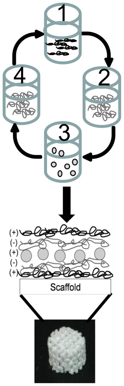
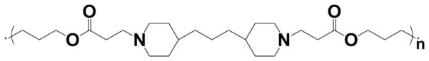
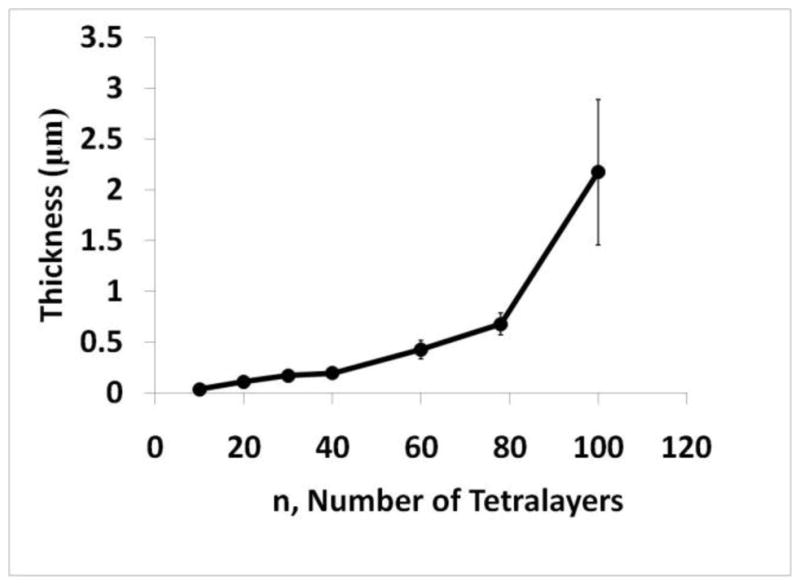
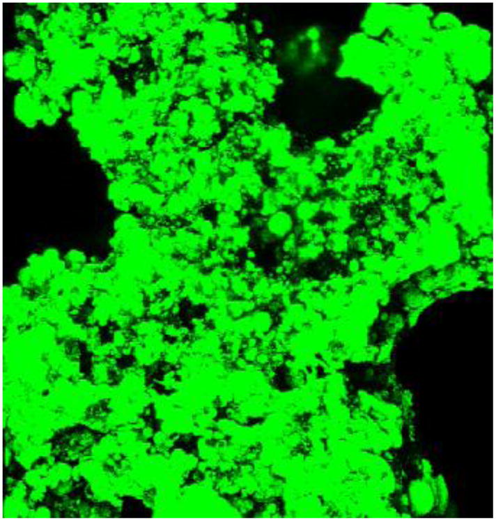
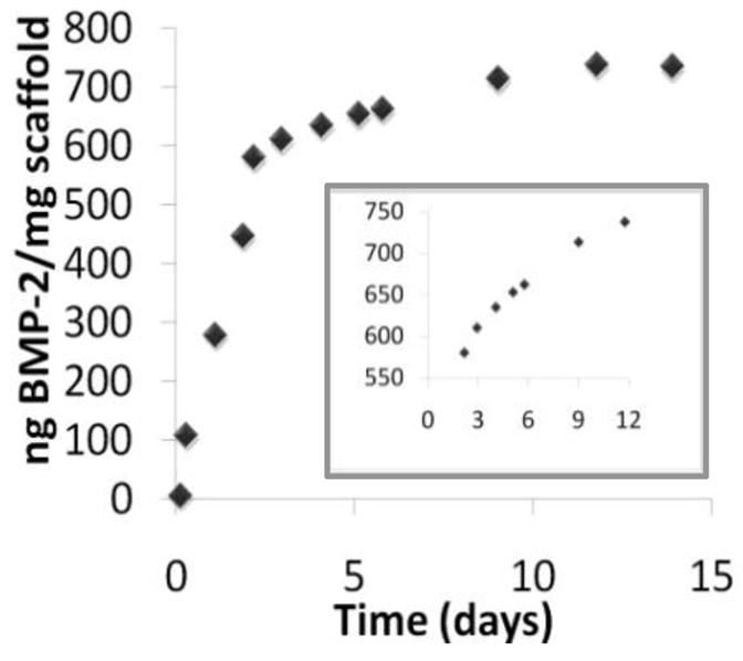
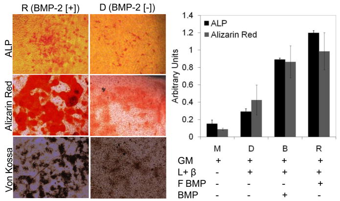
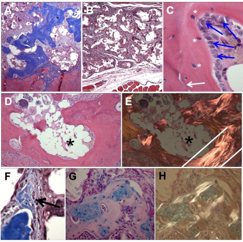
Drug eluting coatings that can direct the host tissue response to implanted medical devices have the potential to ameliorate both the medical and financial burden of complications from implantation. However, because many drugs useful in this arena are biologic in nature, a paucity of delivery strategies for biologics, including growth factors, currently limits the control that can be exerted on the implantation environment. Layer-by-Layer (LbL) polyelectrolyte multilayer films are highly attractive as ultrathin biologic reservoirs, due to the capability to conformally coat difficult geometries, the use of aqueous processing likely to preserve fragile protein function, and the tunability of incorporation and release profiles. Herein, we describe the first LbL films capable of microgram-scale release of the biologic Bone Morphogenetic Protein 2 (BMP-2), which is capable of directing the host tissue response to create bone from native progenitor cells. Ten micrograms of BMP-2 are released over a period of two weeks in vitro; less than 1% is released in the first 3 h (compared with commercial collagen matrices which can release up to 60% of BMP-2, too quickly to induce differentiation). BMP-2 released from LbL films retains its ability to induce bone differentiation in MC3T3 E1S4 pre-osteoblasts, as measured by induction of alkaline phosphatase and stains for calcium (via Alizarin Red) and calcium matrix (via Von Kossa). In vivo, BMP-2 film coated scaffolds were compared with film coated scaffolds lacking BMP-2. BMP-2 coatings implanted intramuscularly were able to initiate host progenitor cells to differentiate into bone, which matured and expanded from four to 9 weeks as measured by MicroCT and histology. Such LbL films represent new steps towards controlling and tuning host response to implanted medical devices, which may ultimately increase the success of implanted devices, provide alternative new approaches toward bone wound healing, and lay the foundation for development of a multi-therapeutic release coating.


Figures

Figure 1
Figure 1
Schematic of LbL architecture shows that a 3DP scaffold (or glass surface) is repeatedly dipped with tetralayer units consisting of (1) Poly 2 (positively charged), (2) chondroitin sulphate (negatively charged), (3) BMP-2 (positively charged) and (4) chondroitin sulfate. This tetralayer structure is repeated 100 times for all LbL films in this communication.
Figure 2
Figure 2
Structure of Poly2, the selected poly(-aminoester) used in this work.
Figure 3
Figure 3
Profilometry measurements of LbL films built on glass allow for thickness analysis. Film thickness grows exponentially (R2 = 0.96) with increasing numbers of tetralayers in a [P2/Chondroitin/BMP-2/Chondroitin]n film.
Figure 4
Figure 4
A 3DP βTCP-PCL blend scaffold was dipped with [P2/chondroitin/fluorescent lysozyme/chondroitin]100. A three dimensional reconstruction of the resulting fluorescent LbL film shows a coating with conforms to the roughened, popcorn ball-like surface of the underlying 3DP scaffold.
Figure 5
Figure 5
Release of BMP-2 from LbL films built on 3DP scaffold. Eighty percent of the material is released over a two day, linear release period. The remaining twenty percent is released over a period of approximately two weeks. Inset shows a blow up of the release profile after the initial two day period. Fourteen mg of scaffold were inserted in in vivo experiments; thus approximately 10 ug of BMP-2 were released from scaffolds.
Figure 6
Figure 6
Left panel: Bioactive BMP-2 released from LbL films (sample R) induces differentiation of MC3T3 pre-osteoblasts to an osteoblast phenotype compared with differentiation medium alone (sample D). Alkaline Phosphatase (ALP) staining (red) shows early (day 6) activation of the bone differentiation cascade; At 28 days of culture Alizarin Red staining for calcium deposition (red), and Von Kossa staining for mineralization of the calcium matrix (black) confirm the differentiation process to osteoblasts. Right panel: Quantification of the staining signal (3 readings on four independent experiments; error bar is standard deviation) shows that the increase in activity of BMP-2 released from LbL films (R) over BMP-2 positive control (B)(matched 90 ng/mL for both samples) is statistically significant. A single factor ANOVA test allowed rejection of the null hypothesis for both assays; a Tukey test showed that B and R are statistically different with p<0.01 for ALP assay. Both B and R outperform differentiation medium alone (D) or growth medium alone(M). GM = growth medium; L+β = L-ascorbic acid and β-glycerol phosphate (differentiation factors); F BMP = LbL Film-released BMP-2; BMP = BMP-2 added directly to the medium.
Figure 7
Figure 7
Host progenitor cells exposed to LbL BMP-2 films undergo bone induction, creating calcium deposits visible with microCT in an intramuscular site. MicroCT 2-dimensional slices through the implant (top left panels) show that bone plate thickness is increasing with time; three-dimensional reconstructions (top middle panels) show remarkable pattern fidelity to the underlying scaffold, suggesting that bone grows in an outward-radiating pattern. Control films lacking BMP-2 show very little radio opacity (top right panels) indicating that BMP-2 is necessary for bone induction; three-dimensional images could not be reconstructed. In the bottom graph, bone mineral density, a marker of bone maturity, increases with implant age, indicating continual remodelling and maturing within the bone tissue. Scalebars are 4 mm.
Figure 8
Figure 8
Histologic findings from in vivo studies.. (A and B) Active scaffolds coated with BMP-2, Poly2, chondroitin sulfate film (A) and control scaffolds coated with Poly2 and chondroitin sulfate film (B), both at 9 weeks post-implantation. Brilliant blue staining of lamellar bone is seen in the active, but not control scaffolds (Masson trichrome stain, 40× original magnification). (C) Lamellar bone showing active bone formation with osteoblasts (blue arrows), mature osteocytes in lacunae (white asterisks) and cement line (white arrow) in active scaffolds at 9 weeks post-implantation. (H&E staining, 400× original magnification). (D and E) Mature lamellar bone with fatty marrow space (asterisk) under normal (D) and polarized light microscopy (E) at 9 weeks post-implantation. Note the bright signal from parallel collagen fibers indicating highly aligned lamellar bone, especially between the white lines. (H&E staining, 100× original magnification) (F) Woven bone formation (arrow) seen as a focus with pale blue staining and higher cellularity (black arrow) in contrast to the mature lamellar bone just below at 4 weeks post-implantation (Masson trichrome stain, 100× original magnification). (G and H) Cartilaginous intermediate areas (blue staining in G) are being remodeled into mature bone at 4 weeks post-implantation. The more mature areas brightly polarize while the immature cartilaginous areas do not (in H) (Alcian blue staining, 200× original magnification).

References

    1. Sheridan C. Fresh from the biologic pipeline-2009. Nat Biotechnol. 2009;28(4):307–310. - PubMed
    1. Lavernia CJ, Drakeford MK, Tsao AK, Gittelsohn A, Krackow KA, Hungerford DS. Revision and primary hip and knee arthroplasty - a cost-analysis. Clin Orthop Relat Res. 1995;311:136–141. - PubMed
    1. Fu K, Pack DW, Klibanov AM, Langer R. Visual evidence of acidic environment within degrading poly(lactic-co-glycolic acid) (PLGA) microspheres. Pharm Res. 2000;17(1):100–106. - PubMed
    1. Jewell CM, Lynn DM. Multilayered polyelectrolyte assemblies as platforms for the delivery of DNA and other nucleic acid-based therapeutics. Adv Drug Deliv Rev. 2008;60(9):979–999. - PMC - PubMed
    1. Schneider A, Vodouhe C, Richert L, Francius G, Le Guen E, Schaaf P, et al. Multifunctional polyelectrolyte multilayer films: Combining mechanical resistance, biodegradability, and bioactivity. Biomacromolecules. 2007;8(1):139–145. - PMC - PubMed

Publication types

Substances