

The BH3 mimetic ABT-737 induces cancer cell senescence
- PMID: 21084274
- PMCID: PMC3459262
- DOI: 10.1158/0008-5472.CAN-10-1977
The BH3 mimetic ABT-737 induces cancer cell senescence
Abstract
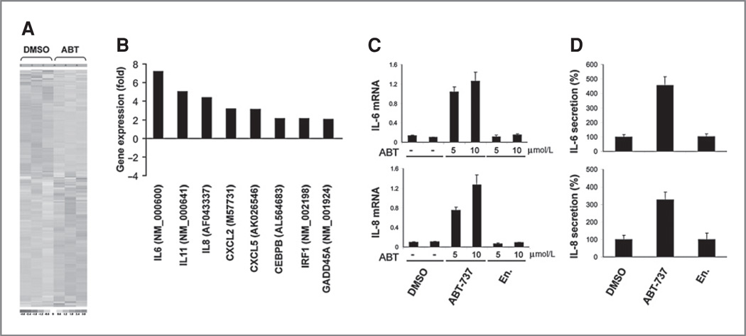
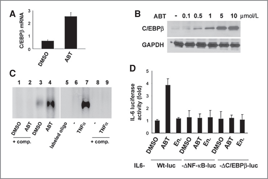
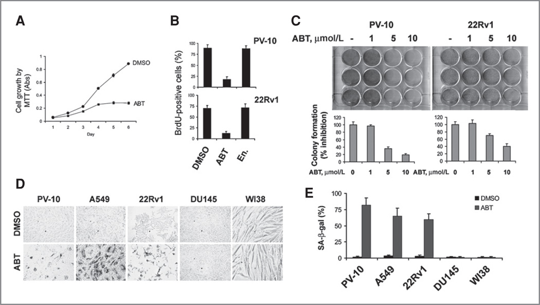
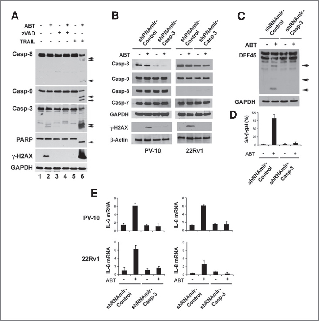
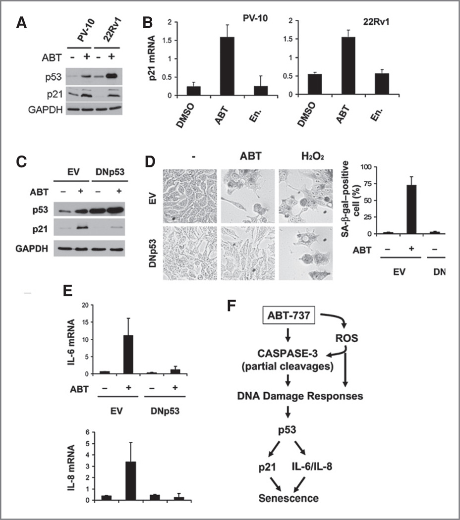
ABT-737, a small molecule cell-permeable Bcl-2 antagonist that acts by mimicking BH3 proteins, induces apoptotic cell death in multiple cancer types. However, when incubated with this agent many solid tumor cell lines do not undergo apoptosis. The current study reveals a novel mechanism whereby ABT-737 when added to apoptosis-resistant cancer cells has profound biologic effects. In PV-10 cells, a renal cell carcinoma that does not die after ABT-737 treatment, this agent induces a two-fold change in the transcription of nearly 430 genes. Many of these induced mRNA changes are in secreted proteins, IL-6, IL-8, and IL-11 and chemokines CXCL2 and CXCL5, or genes associated with an "inflammatory" phenotype. Strikingly, these gene changes are highly similar to those changes previously identified in cellular senescence. Brief exposure of apoptosis-resistant renal, lung and prostate cancer cell lines to ABT-737, although not capable of inducing cell death, causes the induction of senescence-associated β-galactosidase and inhibition of cell growth consistent with the induction of cellular senescence. Evidence indicates that the induction of senescence occurs as a result of reactive oxygen species elevation followed by low-level activation of the caspase cascade, insufficient to induce apoptosis, but sufficient to lead to minor DNA damage and increases in p53, p21, IL-6 and 8 proteins. By overexpression of a dominant-negative p53 protein, we show that ABT-737-induced cellular senescence is p53-dependent. Thus, in multiple cancer types in which ABT-737 is incapable of causing cell death, ABT-737 may have additional cellular activities that make its use as an anticancer agent highly attractive.
© 2010 AACR.
Conflict of interest statement
No potential conflicts of interests were disclosed.
Figures


Similar articles
-
The combination of BH3-mimetic ABT-737 with the alkylating agent temozolomide induces strong synergistic killing of melanoma cells independent of p53.PLoS One. 2011;6(8):e24294. doi: 10.1371/journal.pone.0024294. Epub 2011 Aug 29. PLoS One. 2011. PMID: 21897876 Free PMC article.
-
ABT-737 induces expression of the death receptor 5 and sensitizes human cancer cells to TRAIL-induced apoptosis.J Biol Chem. 2008 Sep 5;283(36):25003-13. doi: 10.1074/jbc.M802511200. Epub 2008 Jul 3. J Biol Chem. 2008. PMID: 18599488 Free PMC article.
-
Cucurbitacin-I inhibits Aurora kinase A, Aurora kinase B and survivin, induces defects in cell cycle progression and promotes ABT-737-induced cell death in a caspase-independent manner in malignant human glioma cells.Cancer Biol Ther. 2015;16(2):233-43. doi: 10.4161/15384047.2014.987548. Cancer Biol Ther. 2015. PMID: 25482928 Free PMC article.
-
Small molecule inhibition of the Bcl-X(L)-BH3 protein-protein interaction: proof-of-concept of an in vivo chemopotentiator ABT-737.Curr Top Med Chem. 2007;7(10):961-5. doi: 10.2174/156802607780906843. Curr Top Med Chem. 2007. PMID: 17508927 Review.
-
Targeting multiple arms of the apoptotic regulatory machinery.Cancer Res. 2007 Apr 1;67(7):2908-11. doi: 10.1158/0008-5472.CAN-07-0082. Cancer Res. 2007. PMID: 17409392 Review.
Cited by
-
Increased Bcl-xL Expression in Pancreatic Neoplasia Promotes Carcinogenesis by Inhibiting Senescence and Apoptosis.Cell Mol Gastroenterol Hepatol. 2017 Feb 20;4(1):185-200.e1. doi: 10.1016/j.jcmgh.2017.02.001. eCollection 2017 Jul. Cell Mol Gastroenterol Hepatol. 2017. PMID: 28948203 Free PMC article.
-
Apoptosis - Fueling the oncogenic fire.FEBS J. 2021 Aug;288(15):4445-4463. doi: 10.1111/febs.15624. Epub 2020 Nov 25. FEBS J. 2021. PMID: 33179432 Free PMC article. Review.
-
ABT-737 synergizes with Bortezomib to kill melanoma cells.Biol Open. 2012 Feb 15;1(2):92-100. doi: 10.1242/bio.2011035. Epub 2011 Nov 16. Biol Open. 2012. PMID: 23213401 Free PMC article.
-
Elimination of quiescent/slow-proliferating cancer stem cells by Bcl-XL inhibition in non-small cell lung cancer.Cell Death Differ. 2014 Dec;21(12):1877-88. doi: 10.1038/cdd.2014.105. Epub 2014 Jul 18. Cell Death Differ. 2014. PMID: 25034785 Free PMC article.
-
Sunitinib induces cellular senescence via p53/Dec1 activation in renal cell carcinoma cells.Cancer Sci. 2013 Aug;104(8):1052-61. doi: 10.1111/cas.12176. Epub 2013 May 16. Cancer Sci. 2013. PMID: 23578198 Free PMC article.
References
-
- Oltersdorf T, Elmore SW, Shoemaker AR, Armstrong RC, Augeri DJ, Belli BA, et al. An inhibitor of Bcl-2 family proteins induces regression of solid tumours. Nature. 2005;435:677–681. - PubMed
-
- Chauhan D, Velankar M, Brahmandam M, Hideshima T, Podar K, Richardson P, et al. A novel Bcl-2/Bcl-X(L)/Bcl-w inhibitor ABT-737 as therapy in multiple myeloma. Oncogene. 2007;26:2374–2380. - PubMed
-
- Tagscherer KE, Fassl A, Campos B, Farhadi M, Kraemer A, Böck BC, et al. Apoptosis-based treatment of glioblastomas with ABT-737, a novel small molecule inhibitor of Bcl-2 family proteins. Oncogene. 2008;27:6646–6656. - PubMed
-
- Tahir SK, Yang X, Anderson MG, Morgan-Lappe SE, Sarthy AV, Chen J, et al. Influence of Bcl-2 family members on the cellular response of small-cell lung cancer cell lines to ABT-737. Cancer Res. 2007;67:1176–1183. - PubMed
Publication types
MeSH terms
Substances
Grants and funding
LinkOut - more resources
Full Text Sources
Other Literature Sources
Research Materials
Miscellaneous
