. 2011 Jan 28;286(4):2987-97.
doi: 10.1074/jbc.M110.190538. Epub 2010 Nov 17.

Structural and biophysical analysis of BST-2/tetherin ectodomains reveals an evolutionary conserved design to inhibit virus release

Affiliations

Structural and biophysical analysis of BST-2/tetherin ectodomains reveals an evolutionary conserved design to inhibit virus release

Melissa Swiecki et al. J Biol Chem. .

Abstract

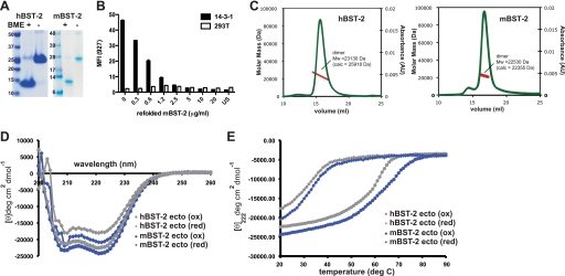
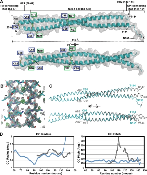
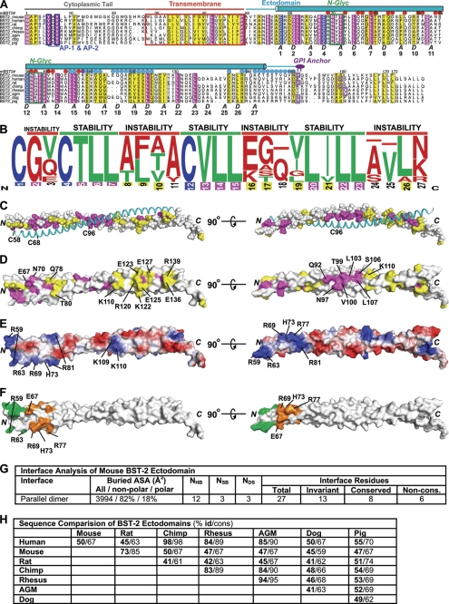
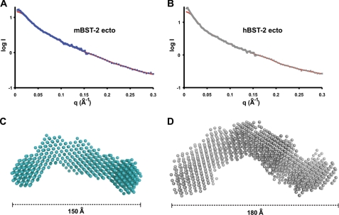
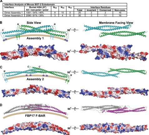
BST-2/tetherin is a host antiviral molecule that functions to potently inhibit the release of enveloped viruses from infected cells. In return, viruses have evolved antagonists to this activity. BST-2 traps budding virions by using two separate membrane-anchoring regions that simultaneously incorporate into the host and viral membranes. Here, we detailed the structural and biophysical properties of the full-length BST-2 ectodomain, which spans the two membrane anchors. The 1.6-Å crystal structure of the complete mouse BST-2 ectodomain reveals an ∼145-Å parallel dimer in an extended α-helix conformation that predominantly forms a coiled coil bridged by three intermolecular disulfides that are required for stability. Sequence analysis in the context of the structure revealed an evolutionarily conserved design that destabilizes the coiled coil, resulting in a labile superstructure, as evidenced by solution x-ray scattering displaying bent conformations spanning 150 and 180 Å for the mouse and human BST-2 ectodomains, respectively. Additionally, crystal packing analysis revealed possible curvature-sensing tetrameric structures that may aid in proper placement of BST-2 during the genesis of viral progeny. Overall, this extended coiled-coil structure with inherent plasticity is undoubtedly necessary to accommodate the dynamics of viral budding while ensuring separation of the anchors.


Figures

FIGURE 1.
FIGURE 1.
Biochemical and biophysical properties of BST-2 ectodomains. A, refolded BST-2 ectodomains are disulfide-linked covalent dimers. SDS-PAGE of the hBST-2 and mBST-2 ectodomains was performed with and without reducing agent (β-mercaptoethanol (BME)). B, the refolded mBST-2 ectodomain competes with BST-2-expressing 14-3-1 cells for binding to anti-mBST-2 mAb 927. The bar graph shows the mean fluorescence intensity (MFI) of mAb 927 staining using increasing concentrations of mBST-2. HEK293T cells (which do not express BST-2) were used as a negative control. C, MALS of the mBST-2 (right panel) and hBST-2 (left panel) ectodomains. The molecular mass determined closely matched that of the covalent dimer in each case (for hBST-2, MALS and actual molecular masses = 23,130 and 25,918 Da, respectively; and for mBST-2, MALS and actual molecular masses = 22,530 and 22,355, Da, respectively). AU, absorbance units. D, CD spectra of the hBST-2 and mBST-2 ectodomains (ecto) under native (oxidized (ox)) and reduced (red) conditions. Spectra are plotted as mean ellipticity per residue. deg, degrees. E, thermal denaturation by measurement of CD at 222 nm for the hBST-2 and mBST-2 ectodomains under native and reduced conditions. Both showed a dramatic reduction in denaturation temperature when the disulfide bonds were broken. The calculated Tm values are as follows: for mBST-2, 68 °C (oxidized) and 32 °C (reduced); and for hBST-2, 61 °C (oxidized) and 31 °C (reduced).
FIGURE 2.
FIGURE 2.
Crystal structure of the mBST-2 ectodomain (amino acids 53–151). A, the 1.6-Å crystal structure of the mBST-2 ectodomain reveals a parallel α-helical covalent dimer bridged by three intermolecular disulfide bonds. The approximate dimensions shown were calculated in MOLEMAN2. Chain A includes residues 57–151, whereas chain B includes residues 58–144. Cysteine residues involved in intermolecular disulfides (blue boxes) are shown as sticks, as are the two N-linked glycosylation sites (green boxes). TM, transmembrane region. B, electron density after rigid body refinement of the molecular replacement solution. The model shown is the final coordinates. Grey mesh represents a 2FoFc map contoured at 2.0 σ, whereas green mesh represents FoFc contoured at 3.0 σ. Note the readily identifiable disulfide electron density. C, superposition of the mBST-2 ectodomain (cyan) and an hBST-2 ectodomain fragment (grey; Protein Data Bank code 2X7A) (32). Structures were superposed using the “user defined match” option in CCP4MG Version 2.4.0. The Cα r.m.s.d. between superposed dimers is 2.35 Å (r.m.s.d. for individual chains are 1.15 and 1.10 Å, respectively). D, comparison of coiled-coil (CC) parameters for the mBST-2 (blue) and hBST-2 (grey) ectodomains. Values were measured using TWISTER (42). Ang., angstrom.
FIGURE 3.
FIGURE 3.
Sequence and surface analysis of the BST-2 ectodomain. A, alignment of BST-2 sequences. Invariant residues (except cysteines) are magenta, invariant cysteines are blue, and residues highlighted yellow denote a conservation index of 6 or higher as determined by ALSCRIPT (43). The secondary structure of the mBST-2 ectodomain is shown above the alignment. Numbering is from mBST-2 (mBST-2#). Green boxes denote the N-linked glycosylation (N-Glyc) sites, and blue boxes highlight trafficking motifs utilized by AP-1 and AP-2. GPI anchor positions predicted by the BIG-PI Predictor (44) are shown as purple boxes. The letters A and D indicate coiled-coil core heptad repeat positions as determined by TWISTER. Dots above the sequence indicate residues located at the interface of the two possible dimer-of-dimers assemblies in the crystal: assembly 1 (red dots), assembly 2 (blue dots), and both assemblies (violet dots). Suffixes in sequence names indicate species as follows: mouse, Mus musculus; human, Homo sapiens; rat, Rattus norvegicus; chimp, Pan troglodytes; rhesus, Macaca mulatta; African green monkey (agm), Chlorocebus tantalus; dog, Canis lupus familiaris; and pig, Sus scrofa. B, sequence logo frequency plot of coiled-coil interface residues. Blue, cysteine; green, favorable coiled-coil interface residues (ILVT); red, unfavorable coiled-coil interface residues (all others). Numbering of interface position is color-coded according to sequence conservation as shown above. C, sequence conservation and invariance at the covalent dimer interface. One chain of the dimer is shown as space-filled and colored according to the sequence alignment shown in A, whereas the other is shown as a cyan coil. D, sequence conservation mapped onto the solvent-accessible surface of the mBST-2 ectodomain. E, charge-smoothened electrostatic surface as calculated by PyMOL. Blue denotes positively charged surfaces, whereas red denotes negatively charged surfaces (gradient, +70 to −70). F, surface mapping of cluster mutations previously demonstrated (32) that do not (green) and do (orange) prevent hBST-2 from restricting HIV-1 budding. These residues (mouse/human) are Arg-59/Arg-54, Asp-60/Asp-55, and Arg-63/Arg-58 (green) and Glu-67/Glu-62, Arg-69/Arg-64, Asn-70/Asn-65, His-73/His-68, Gln-76/Gln-71, Arg-77/Gln-72, and Gln-78/Glu-73 (orange). G, interface analysis of the mBST-2 ectodomain covalent dimer. ASA, accessible surface area; NHB, number of hydrogen bonds; NSB, number of salt bridges; NDS, number of disulfide bonds; Non-cons., non-conserved. H, table of direct sequence comparisons between BST-2 ectodomains from various species.
FIGURE 4.
FIGURE 4.
Solution SAXS of BST-2 ectodomains. A and B, experimental scattering for the mBST-2 ectodomain (ecto) at 5 mg/ml (blue ×) and the hBST-2 ectodomain at 5 mg/ml (grey ×), respectively, along with calculated scattering (red lines) from ab initio models with lowest χ values. Experimental scattering is the average of three experiments with subtraction of buffer scattering. C and D, ab initio models of mBST-2 and hBST-2 ectodomains, respectively, indicate bent flexible structures in solution. Models are the average of 10 independent GASBOR predictions averaged in DAMAVER.
FIGURE 5.
FIGURE 5.
Possible assemblies created by crystal packing. A, interface surface analysis for the two major assemblies created by crystal packing. Symmetry operations creating the assemblies were as follows: assembly 1, −x + 5/2, y, −z + 1; and assembly 2, −x + 1/2, y, −z. ASA, accessible surface area; NHB, number of hydrogen bonds; NSB, number of salt bridges; NDS, number of disulfide bonds; Non-cons., non-conserved. B and C, assemblies 1 and 2, respectively, viewed from the perspective of a side view on an outwardly curving membrane (left) and the membrane-facing surface (right). Below the ribbon diagrams are the respective electrostatic surfaces as calculated by PyMOL. For B, the C-terminal GPI-connecting loop has been removed from chain A. D, comparison with the FBP17 F-BAR domain (Protein Data Bank code 2EFL), a member of the BAR domain superfamily that binds to curved membranes.

Similar articles

Cited by

References

    1. Wolf D., Goff S. P. (2008) Annu. Rev. Genet. 42, 143–163 - PMC - PubMed
    1. Sadler A. J., Williams B. R. (2008) Nat. Rev. Immunol. 8, 559–568 - PMC - PubMed
    1. Sauter D., Specht A., Kirchhoff F. (2010) Cell 141, 392–398 - PubMed
    1. Evans D. T., Serra-Moreno R., Singh R. K., Guatelli J. C. (2010) Trends Microbiol. 18, 388–396 - PMC - PubMed
    1. Douglas J. L., Gustin J. K., Viswanathan K., Mansouri M., Moses A. V., Früh K. (2010) PLoS Pathog. 6, e1000913. - PMC - PubMed

MeSH terms

Associated data

LinkOut - more resources