

Somatic cell plasticity and Niemann-Pick type C2 protein: fibroblast activation
- PMID: 21084287
- PMCID: PMC3023505
- DOI: 10.1074/jbc.M110.135897
Somatic cell plasticity and Niemann-Pick type C2 protein: fibroblast activation
Abstract
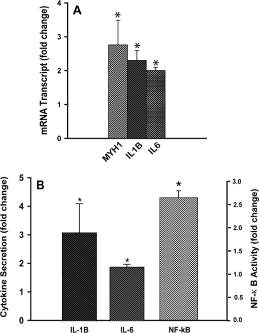
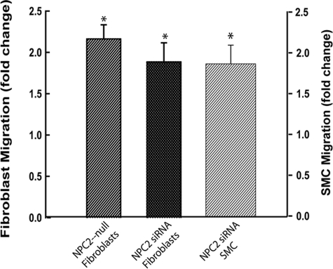
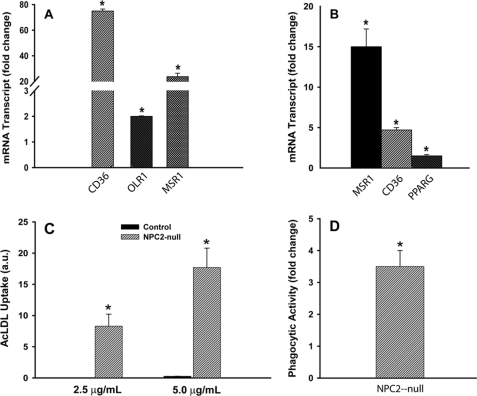
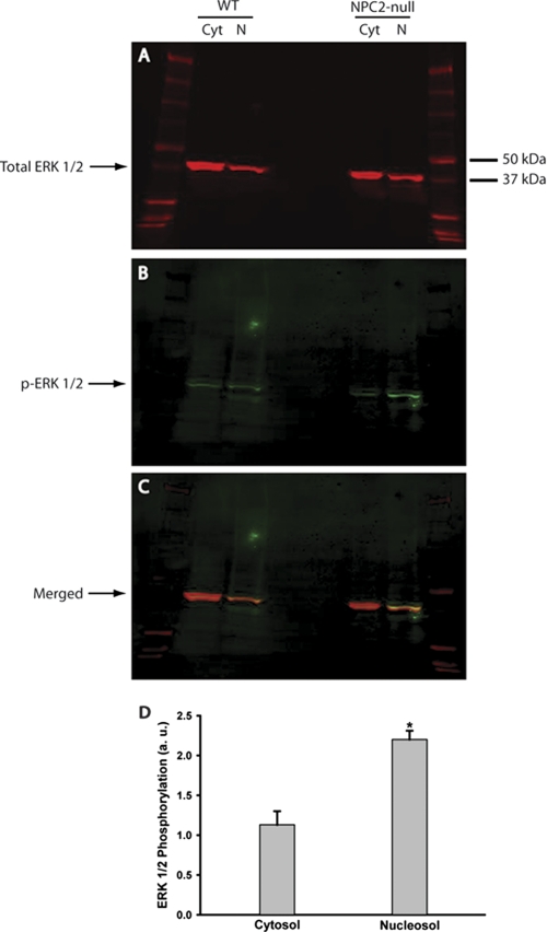
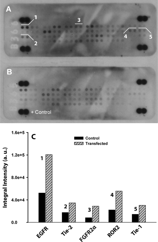
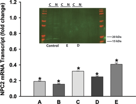
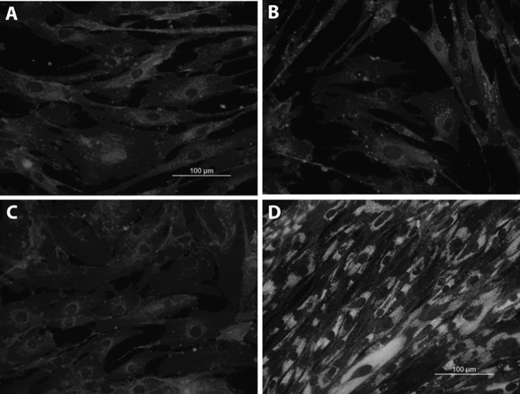
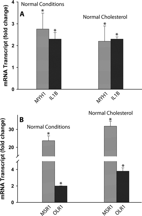
A growing body of evidence points toward activated fibroblasts, also known as myofibroblasts, as one of the leading mediators in several major human pathologies including proliferative fibrotic disorders, invasive tumor growth, rheumatoid arthritis, and atherosclerosis. Niemann-Pick Type C2 (NPC2) protein has been recently identified as a product of the second gene in NPC disease. It encodes ubiquitous, highly conserved, secretory protein with the poorly defined function. Here we show that NPC2 deficiency in human fibroblasts confers their activation. The activation phenomenon was not limited to fibroblasts as it was also observed in aortic smooth muscle cells upon silencing NPC2 gene by siRNA. More importantly, activated synovial fibroblasts isolated from patients with rheumatoid arthritis were also identified as NPC2-deficient at both the NPC2 mRNA and protein levels. The molecular mechanism responsible for activation of NPC2-null cells was shown to be a sustained phosphorylation of ERK 1/2 mitogen-activated protein kinase (MAPK), which fulfills both the sufficient and necessary fibroblast activation criteria. All of these findings highlight a novel mechanism where NPC2 by negatively regulating ERK 1/2 MAPK phosphorylation may efficiently suppress development of maladaptive tissue remodeling and inflammation.
Figures
Similar articles
-
Niemann-pick type C2 deficiency in human fibroblasts confers robust and selective activation of prostaglandin E2 biosynthesis.J Biol Chem. 2013 Aug 16;288(33):23696-703. doi: 10.1074/jbc.M112.445916. Epub 2013 Jun 27. J Biol Chem. 2013. PMID: 23814065 Free PMC article.
-
Somatic cell plasticity and Niemann-pick type C2 protein: adipocyte differentiation and function.J Biol Chem. 2010 Sep 24;285(39):30347-54. doi: 10.1074/jbc.M110.135939. Epub 2010 Jul 22. J Biol Chem. 2010. PMID: 20650896 Free PMC article.
-
Multiple Surface Regions on the Niemann-Pick C2 Protein Facilitate Intracellular Cholesterol Transport.J Biol Chem. 2015 Nov 6;290(45):27321-27331. doi: 10.1074/jbc.M115.667469. Epub 2015 Aug 20. J Biol Chem. 2015. PMID: 26296895 Free PMC article.
-
Do GWAS and studies of heterozygotes for NPC1 and/or NPC2 explain why NPC disease cases are so rare?J Appl Genet. 2018 Nov;59(4):441-447. doi: 10.1007/s13353-018-0465-2. Epub 2018 Sep 13. J Appl Genet. 2018. PMID: 30209687 Review.
-
Niemann-Pick C2 (NPC2) and intracellular cholesterol trafficking.Biochim Biophys Acta. 2009 Jul;1791(7):671-8. doi: 10.1016/j.bbalip.2009.02.001. Epub 2009 Feb 13. Biochim Biophys Acta. 2009. PMID: 19232397 Free PMC article. Review.
Cited by
-
Protein replacement therapy partially corrects the cholesterol-storage phenotype in a mouse model of Niemann-Pick type C2 disease.PLoS One. 2011;6(11):e27287. doi: 10.1371/journal.pone.0027287. Epub 2011 Nov 3. PLoS One. 2011. PMID: 22073306 Free PMC article.
-
Systemic Investigation of Promoter-wide Methylome and Genome Variations in Gout.Int J Mol Sci. 2020 Jul 1;21(13):4702. doi: 10.3390/ijms21134702. Int J Mol Sci. 2020. PMID: 32630231 Free PMC article.
-
Niemann-pick type C2 deficiency in human fibroblasts confers robust and selective activation of prostaglandin E2 biosynthesis.J Biol Chem. 2013 Aug 16;288(33):23696-703. doi: 10.1074/jbc.M112.445916. Epub 2013 Jun 27. J Biol Chem. 2013. PMID: 23814065 Free PMC article.
-
Characterization of Niemann-Pick Type C2 protein expression in multiple cancers using a novel NPC2 monoclonal antibody.PLoS One. 2013 Oct 17;8(10):e77586. doi: 10.1371/journal.pone.0077586. eCollection 2013. PLoS One. 2013. PMID: 24147030 Free PMC article.
-
Changes on the Caco-2 secretome through differentiation analyzed by 2-D differential in-gel electrophoresis (DIGE).Int J Mol Sci. 2012 Nov 7;13(11):14401-20. doi: 10.3390/ijms131114401. Int J Mol Sci. 2012. PMID: 23203071 Free PMC article.
References
Publication types
MeSH terms
Substances
Grants and funding
LinkOut - more resources
Full Text Sources
Miscellaneous
