. 2011 Jan 14;286(2):1227-36.
doi: 10.1074/jbc.M110.194613. Epub 2010 Nov 17.

Dematin, a component of the erythrocyte membrane skeleton, is internalized by the malaria parasite and associates with Plasmodium 14-3-3

Affiliations

Dematin, a component of the erythrocyte membrane skeleton, is internalized by the malaria parasite and associates with Plasmodium 14-3-3

Marco Lalle et al. J Biol Chem. .

Abstract

The malaria parasite invades the terminally differentiated erythrocytes, where it grows and multiplies surrounded by a parasitophorous vacuole. Plasmodium blood stages translocate newly synthesized proteins outside the parasitophorous vacuole and direct them to various erythrocyte compartments, including the cytoskeleton and the plasma membrane. Here, we show that the remodeling of the host cell directed by the parasite also includes the recruitment of dematin, an actin-binding protein of the erythrocyte membrane skeleton and its repositioning to the parasite. Internalized dematin was found associated with Plasmodium 14-3-3, which belongs to a family of conserved multitask molecules. We also show that, in vitro, the dematin-14-3-3 interaction is strictly dependent on phosphorylation of dematin at Ser(124) and Ser(333), belonging to two 14-3-3 putative binding motifs. This study is the first report showing that a component of the erythrocyte spectrin-based membrane skeleton is recruited by the malaria parasite following erythrocyte infection.


Figures

FIGURE 1.
FIGURE 1.
Expression and localization of Pb14-3-3 protein. A, equal amounts of protein extracts from mouse (m) and human (h) RBCs, T. gondii tachyzoites, C. parvum sporozoites, and asynchronous P. falciparum and P. berghei parasites were blotted and sequentially probed with mouse α-Pb14-3-3 serum and rabbit α-pan 14-3-3 polyclonal Abs. 1 μg of recombinant Pb14-3-3 (FLAG-Pb14-3-3) was used as a control. Two different exposures of α-pan 14-3-3 are presented. B, soluble proteins from P. berghei trophozoite (Troph.), schizont (Schiz.), and gametocyte (Gamet.) stages were blotted and sequentially probed with α-Pb14-3-3 and α-PfHsp70 immune sera. C, CLSM observations of mouse erythrocytes infected by P. berghei at different developmental stages: free merozoite (panel a), trophozoite (panel b), schizont (panel c), and gametocyte (panel d), stained with α-SEP1 (red) and α-Pb14-3-3 (green). Nuclei are stained with DAPI (blue). Displayed micrographs correspond to a single stack encompassing the center of the nucleus. T, transmission light acquisition. Scale bars, 1 μm.
FIGURE 2.
FIGURE 2.
Erythroid dematin directly interacts with Pb14-3-3. A, glutathione-Sepharose-immobilized GST-Pb14-3-3 or GST as a control was incubated with a P. berghei extract solubilized by sonication. The eluted proteins were subjected to Western blot analysis using an α-dematin mAb. B, dematin was immunopurified from P. berghei parasites using the specific α-dematin mAb. Parasite extract (input, 1/100), pretreated with protein-G beads (preclearing, 1/20), was incubated with α-dematin-conjugated protein-G beads. After removal of the unbound material (flow-through, 1/100), immunoprecipitated proteins were eluted (IP, 1/20). Blotted samples were probed with α-dematin mAb and α-Pb14-3-3. The overlay assay was performed using GST-Pb14-3-3 as a probe. The interactions were inhibited through the addition of the A8Ap phosphopeptide. Asterisks in both panels indicate the bands corresponding to the 48- and 52-kDa dematin.
FIGURE 3.
FIGURE 3.
Subcellular localization of dematin in P. berghei-infected mouse RBC. A, CLSM observations of mouse erythrocytes infected by P. berghei at different developmental stages: trophozoite (panel a), schizont (panel b), gametocyte (panel c), and a normal RBC (panel d), stained with α-SEP1 (red) and α-dematin mAb (green). Nuclei were stained with DAPI (blue). Displayed micrographs correspond to the single stack encompassing the center of the parasite nucleus. T, transmission light acquisition. Scale bars, 0.5 μm. B, soluble (S), high salt soluble (HS), and insoluble (I) proteins from P. berghei young (Troph. 13h) and late (Troph. 17h) trophozoites, schizonts (Schiz.), gametocytes (Gamet.) and total ghost proteins from normal mouse RBCs were blotted and sequentially probed with α-Pb14-3-3 and α-dematin mAb. Arrows indicate the 48–52-kDa dematin doublet. C, P. berghei soluble (S) and insoluble (I) proteins obtained by sonication were blotted and sequentially probed with α-Pb14-3-3 and α-dematin mAb. Scale bars, 1 μm.
FIGURE 4.
FIGURE 4.
Localization of dematin in P. falciparum iRBC. A, ghosts (G) and parasite-enriched fraction (T) were separated from iRBCs. Parasite fraction was treated with Triton X-100 to obtain soluble (S) and insoluble (I) proteins. Blotted samples were probed with α-dematin mAb, α-Pb14-3-3, and α-Hu14-3-3γ serum (specific for the human 14-3-3 isoforms). B, CLSM observations of normal human RBCs (panel a) and erythrocytes infected by P. falciparum at different developmental stages: trophozoite (panel a), schizont (panel b), and gametocyte (panel c), stained with the α-EXP1 serum (green), which decorates the PVM, and the α-dematin md52k serum (red). Nuclei were stained with DAPI (blue). Displayed micrographs correspond to a single stack encompassing the center of the nucleus. T, transmission light acquisition. Scale bars, 1 μm. C, parasite fraction (PF) and ghosts from iRBC (iG) or normal RBC (G) were treated with streptolysin-O and incubated (+) or not (−) with PK, blotted, and probed with α-dematin mAb. The effectiveness of PK treatment was confirmed using the α-BR5 immune serum against the P. falciparum Maurer's cleft transmembrane protein Pfsbp1. In the PK-treated sample, α-BR5 reacts with a 37-kDa polypeptide.
FIGURE 5.
FIGURE 5.
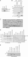
Dematin-Pb14-3-3 interaction is promoted by PKA phosphorylation. A, a GST-fused 52-kDa isoform of dematin was phosphorylated in vitro using the PKA catalytic subunit or casein kinase II (CKII), digested with the PreScission enzyme, and run in SDS-PAGE. A control sample without enzymes was also included. Autoradiography (Autorad.) of the stained gel showed that the recombinant dematin was efficiently labeled only by PKA. B, glutathione-Sepharose-immobilized GST or GST-dematin, phosphorylated (+) or not (−) with PKA, was incubated with a bacterial lysate containing His-Pb14-3-3. Proteins eluted with A8Ap synthetic phosphopeptide or glutathione were blotted and probed with α-His6, to detect the associated 14-3-3, or α-GST, to detect the eluted recombinant GST and GST-dematin. C, putative 14-3-3-binding sites on the 52-kDa dematin isoform are underlined. Putative PKA phosphorylation sites coinciding with the 14-3-3-binding sites are in bold. The position of mutated serines is specified. The ATP-binding site (P-loop) is boxed in gray. D, recombinant GST, GST-dematin, or GST-dematin mutants, bound to glutathione-Sepharose beads, were phosphorylated by the PKA catalytic subunit. Phosphorylated recombinant proteins, digested with the PreScission enzyme, were run in SDS-PAGE, stained with Coomassie Blue, and subjected to autoradiography. E, GST, GST-dematin, and GST-dematin mutants were treated as in panel B. Proteins eluted from the glutathione-Sepharose beads with the A8Ap synthetic phosphopeptide were subjected to immunoblotting using the α-His6 to detect bound 14-3-3.

Similar articles

Cited by

References

    1. Marti M., Good R. T., Rug M., Knuepfer E., Cowman A. F. (2004) Science 306, 1930–1933 - PubMed
    1. Hiller N. L., Bhattacharjee S., van Ooij C., Liolios K., Harrison T., Lopez-Estraño C., Haldar K. (2004) Science 306, 1934–1937 - PubMed
    1. Maier A. G., Cooke B. M., Cowman A. F., Tilley L. (2009) Nat. Rev. Microbiol. 7, 341–354 - PubMed
    1. Azim A. C., Knoll J. H., Beggs A. H., Chishti A. H. (1995) J. Biol. Chem. 270, 17407–17413 - PubMed
    1. Bruce L. J., Beckmann R., Ribeiro M. L., Peters L. L., Chasis J. A., Delaunay J., Mohandas N., Anstee D. J., Tanner M. J. (2003) Blood 101, 4180–4188 - PubMed

Publication types

MeSH terms