. 2011 Jan;25(1):44-57.
doi: 10.1210/me.2010-0249. Epub 2010 Nov 17.

A new, lineage specific, autoup-regulation mechanism for human glucocorticoid receptor gene expression in 697 pre-B-acute lymphoblastic leukemia cells

Affiliations

A new, lineage specific, autoup-regulation mechanism for human glucocorticoid receptor gene expression in 697 pre-B-acute lymphoblastic leukemia cells

Chuan-dong Geng et al. Mol Endocrinol. 2011 Jan.

Abstract

Glucocorticoid (GC) steroid hormones induce apoptosis in acute lymphoblastic leukemia (ALL). Autoup-regulation of human GC receptor (hGR) levels is associated with sensitivity to GC-mediated apoptosis. Among the major hGR promoters expressed in 697 pre-B-ALL cells (1A, 1B, 1C, and 1D), only promoters 1C and 1D are selectively activated by the hormone. Promoter 1B is unresponsive, and promoter 1A is down-regulated by dexamethasone (Dex) in 697 cells, whereas they are both up-regulated in CEM-C7 T-ALL cells. Autoup-regulation of promoter 1C and 1D in 697 cells requires sequences containing GC response units (GRUs) (1C GRU, -2915/-2956; 1D GRU, -4525/-4559) that were identified previously in CEM-C7 cells. These GRUs potentially bind GR, c-myeloblastosis (c-Myb), and E-twenty six (Ets) proteins; 697 cells express high levels of c-Myb protein, as well as the E-twenty six family protein members, PU.1 and Spi-B. Dex treatment in 697 cells elevates the expression of c-Myb and decreases levels of both Spi-B and PU.1. Chromatin immunoprecipitation assays revealed the specific recruitment of GR, c-Myb, and cAMP response element-binding protein binding protein to the 1C and 1D GRUs upon Dex treatment, correlating to observed autoup-regulated activity in these two promoters. These data suggest a hormone activated, lineage-specific mechanism to control the autoup-regulation of hGR gene expression in 697 pre-B-ALL cells via steroid-mediated changes in GR coregulator expression. These findings may be helpful in understanding the mechanism that determines the sensitivity of B-ALL leukemia cells to hormone-induced apoptosis.


Figures

Figure 1
Figure 1
Effects of Dex treatment on the expression of GR mRNA transcript variants in 697 pre-B-ALL cell. Transcript-specific primers/probes and real-time qRT-PCR were used to measure the intracellular concentration for each GR gene transcript variant. An exon 8-9α probe set was used to determine all (total) GR transcripts containing exon 9α and that codes for the major GR protein species in these cells, the GRα protein. Dex-responsive expression of each GR mRNA transcript is plotted as percentage of the respective EtOH vehicle control level. Four biological replicates were used for statistical analysis. ***, P < 0.005; and *, P < 0.05 for the level of GR transcripts in the Dex-treated sample compared with the EtOH-treated control.
Figure 2
Figure 2
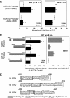
Basal activity and hormone responsiveness of hGR promoters 1C (−2986/−2523) and 1D (−4898/−4525) and their deletions. Human GR promoters 1C and 1D, and their 5′- or 3′-end deletion luciferase reporter gene expression constructs, were transfected into IM-9 or 697 cells along with a β-gal construct (for normalizing transfection efficiency) as described in Materials and Methods. After 24 h of EtOH (vehicle control) or Dex treatment, cell lysates were made, and luciferase assays were performed. A, Basal promoter activity and hormone responsiveness of hGR promoter 1C (−2986/−2523) and promoter 1D (−4898/−4525) in 697 pre-B-ALL and IM-9 B-cells. B, Luciferase reporter gene analysis of promoter 1C and 1D promoter 5′- or 3′-end deletions in 697 pre-B-ALL cells. The results emphasize the important roles of sequences −2915/−2986 of promoter 1C and −4525/−4559 of promoter 1D in both basal promoter activity and hormone response. For A and B, the values are the average of three separate experiments ±sem. *, P < 0.05 for the Dex-induced values vs. the respective EtOH vehicle controls. C, Diagram of the protein binding structure of hormone-responsive sequences located in hGR promoter 1C (−2916/−2956), promoter 1D (−4525/−4559), and promoter 1A (+243/+269).
Figure 3
Figure 3
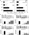
Lineage-specific expression and hormone regulation of transcription factors during hormone-induced apoptosis in 697 pre-B-ALL cells. A, Western blot analysis of the lineage-specific expression of transcription factors in B- and T-lymphoblastic leukemia cells. Total protein lysates from equal numbers of cells were loaded in each SDS-PAGE sample well, followed by electrophoresis, membrane transfer, and blotting with protein-specific antibodies. The display is the scanned x-ray film images. Duplicate samples for each cell line are presented in the top five scans. The sixth series of scans shows that Dex treatment causes the autoup-regulation of GR protein levels in 697 and CEM-C7 ALL cells and the autodown-regulation of GR levels in the IM-9 lymphoblastoid line. Real-time qRT-PCR was used in B–D to examine and compare the lineage-specific expression of transcription factor transcripts and the response to Dex treatment. B, PU.1; C, Spi-B; and D, c-Myb. The results from four or six separate experiments were analyzed statistically. ***, P < 0.005; and **, P < 0.01 for the Dex-treated sample compared with the respective EtOH vehicle control.
Figure 4
Figure 4
Expression of c-Myb in 697 pre-B ALL cells treated with Dex and cytotoxic drugs. A, Time-course (2, 4, 15, and 18 h) expression of c-Myb and total GR mRNA transcripts in 697 cells after Dex treatment. The probes used to measure the GR were from exons 5–6, as described elsewhere (28). B–E, Representative Western blottings of 697 cells treated with Dex or other apoptosis-inducing agents, ara-C, 5-FUDR, and DOXO. The amount of c-Myb protein was determined by densitometry and normalized to the glyceraldehyde-3-phosphate dehydrogenase (GAPDH) protein levels in the same sample. The graphs display the fold differences compared with the control cell samples treated with EtOH or PBS. Treatment times were: B, 2 h; C, 4 h; D, 15 h; and E, 18 h. The experiments were repeated three times and analyzed for statistic significance. *, P < 0.05; **, P < 0.01; and ***, P < 0.005 over vehicle controls.
Figure 5
Figure 5
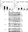
Regulation of the expression of Ets protein family members, PU.1 and Spi-B, by hormone and cytotoxic drugs. The hormone-responsive expression of PU.1 mRNA transcripts (A) and Spi-B (B) was measured in 697 cells after Dex treatment for 2, 4, 15, and 18 h. Three separate experiments were performed. The data show the mean ± sem for percentages in the Dex-treated samples vs. the EtOH-treated control. *, P < 0.05; **, P < 0.01; and ***, P < 0.005. C, Western blot analysis of 697 cells to detect the expression of PU.1 and Spi-B proteins at different times of treatment (2, 4, 15, and 18 h) with Dex or other cytotoxic agents, ara-C, 5-FUDR, and DOXO. GAPDH, glyceraldehyde-3-phosphate dehydrogenase.
Figure 6
Figure 6
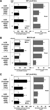
Analysis of c-Myb and PU.1 functions on autoup-regulation of GR 1C and 1D promoters in 697 pre-B-ALL cells. A, A dominant negative inhibitor of c-Myb, the c-Myb DBD, was expressed in 697 cells, and its effect on Dex-mediated autoup-regulation of the hGR 1C- and 1D-luciferase reporter genes was assessed. B, Expression of c-Myb in the IM-9 lymphoblastoid cell (that does not express c-Myb) functionally restored the autoup-regulation of hGR promoters 1C and 1D, as analyzed using luciferase reporter genes. C, Overexpression of PU.1 in 697 cells that blocks the hormone response of the 1D promoter but cannot completely block the Dex induction of 1C promoter, in which the Ets site does not overlap the GR or c-Myb binding sites. For the analysis of each promoter reporter gene construct, the responsiveness to Dex treatment is plotted as the percentage of the respective controls treated with the EtOH vehicle alone (left panels). **, P < 0.01; and *, P < 0.05. The effects of these proteins on basal promoter activity are shown in the right panels.
Figure 7
Figure 7
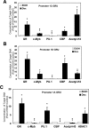
ChIP analysis of in vivo protein binding at hormone-responsive sequences in the hGR 1C, 1D, and 1A promoters in 697 pre-B-ALL cells. Sequence-specific primers/probes (for the hGR 1A, 1C, or 1D GRUs) and real-time qPCR were used to detect and quantify the target genomic DNA sequences that are pulled down by specific protein antibodies via a ChIP assay. The measured quantity of target sequence is normalized to its input amount. The results are plotted as the fold difference in signal obtained in the Dex-treated cells vs. the EtOH vehicle-treated controls to show the increased or decreased binding of each protein to the targeted DNA sequences. Cells were collected for ChIP assay at 17 h after Dex or EtOH addition. The figure shows hormone-altered GR, c-Myb, CBP, PU.1, and HDAC1 (1A promoter only) protein binding, and the relative levels of acetylated histone H3 on the: A, −2916/−2956 1C DNA promoter sequence; B, −4525/−4559 1D DNA promoter sequence; and C, +243/+269 1A DNA promoter sequence. Three separate experiments were used for the statistical analysis. *, P < 0.05; **, P < 0.01; and ***, P < 0.005.
Figure 8
Figure 8
Autoregulation of different GR mRNA transcript variants in Dex-treated primary bone marrow cells from a pre-B-ALL. After in vitro treatment of the purified mononuclear cells with either Dex or the EtOH vehicle, the expression of the various transcripts was determined via qRT-PCR analysis. The value of the Dex-treated sample is expressed as the percentage of that obtained for the respective transcript in the EtOH vehicle-treated control sample.
Figure 9
Figure 9
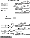
Molecular mechanism for GR autoregulation in pre-B-ALL cells. In the basal state (no steroid), all three promoters (1A, 1C, and 1D) are used to generate GR transcripts. Various transcription factors (some indicated in the figure) are in a dynamic binding equilibrium with GRUs in the GR gene promoters. Upon binding hormone, the GR complex binds to its own promoter and (either directly or indirectly) regulates the transcription rate of various coregulator transcription factors. In human 697 pre-B-ALL cells, the GR down-regulates the expression of the transcription inhibitory Ets proteins, PU.1, and Spi-B, whereas it simultaneously up-regulates its own expression and that of the transcription activator protein, c-Myb. This coordinate down-regulation of transcriptional repressor levels and up-regulation of transcriptional activator proteins causes increase in intracellular GR levels (via the GR 1C and 1D promoters) that can stimulate apoptosis in these cells. The opposite regulation of the hGR 1A promoter under these conditions is not well understood. A possible explanation is presented it the Discussion. GRE, Glucocorticoid response element; EBE1, Ets binding element 1; EBE2, Ets binding element 2.

Similar articles

Cited by

References

    1. Pui CH, Robison LL, Look AT 2008 Acute lymphoblastic leukaemia. Lancet 371:1030–1043 - PubMed
    1. Pui CH 2009 Acute lymphoblastic leukemia: introduction. Semin Hematol 46:1–2 - PMC - PubMed
    1. Nanni P, Nicoletti G, Prodi G, Galli MC, De Giovanni C, Grilli S, Lollini PL, Gobbi M, Cavo M, Tura S 1982 Glucocorticoid receptor and in vitro sensitivity to steroid hormones in human lymphoproliferative diseases and myeloid leukemia. Cancer 49:623–632 - PubMed
    1. Gaynon PS, Carrel, AL 1999 Glucocorticosteroid therapy in childhood acute lymphoblastic leukemia. Adv Exp Med Biol 457:593–605 - PubMed
    1. Pui CH, Evans, WE 2006 Treatment of acute lymphoblastic leukemia. N Engl J Med 354:166–178 - PubMed

Publication types

MeSH terms