. 2011 Jan;152(1):193-206.
doi: 10.1210/en.2010-0802. Epub 2010 Nov 17.

The role of GH and IGF-I in mediating anabolic effects of testosterone on androgen-responsive muscle

Affiliations

The role of GH and IGF-I in mediating anabolic effects of testosterone on androgen-responsive muscle

Carlo Serra et al. Endocrinology. 2011 Jan.

Abstract

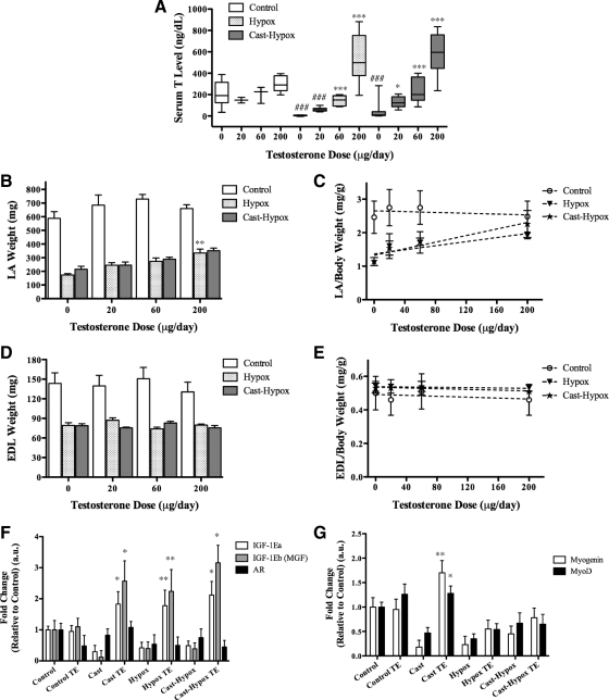
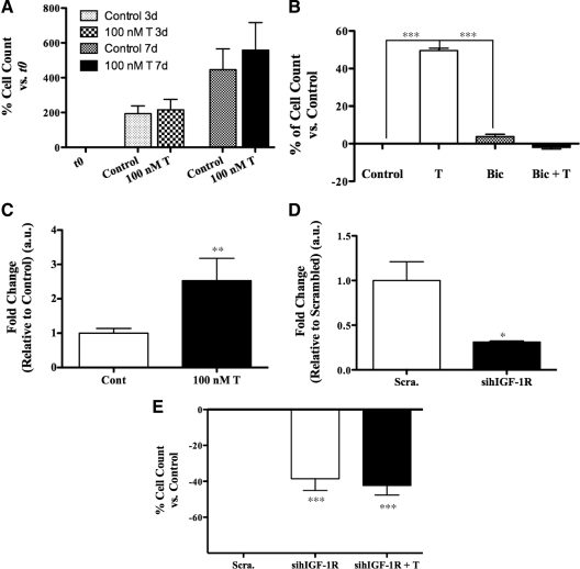
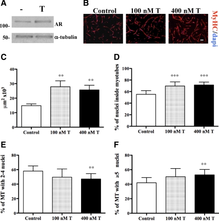
Testosterone (T) supplementation increases skeletal muscle mass, circulating GH, IGF-I, and im IGF-I expression, but the role of GH and IGF-I in mediating T's effects on the skeletal muscle remains poorly understood. Here, we show that T administration increased body weight and the mass of the androgen-dependent levator ani muscle in hypophysectomized as well as castrated plus hypophysectomized adult male rats. T stimulated the proliferation of primary human skeletal muscle cells (hSKMCs) in vitro, an effect blocked by transfecting hSKMCs with small interference RNA targeting human IGF-I receptor (IGF-IR). In differentiation conditions, T promoted the fusion of hSKMCs into larger myotubes, an effect attenuated by small interference RNA targeting human IGF-IR. Notably, MKR mice, which express a dominant negative form of the IGF-IR in skeletal muscle fibers, treated with a GnRH antagonist (acyline) to suppress endogenous T, responded to T administration by an attenuated increase in the levator ani muscle mass. In conclusion, circulating GH and IGF-I are not essential for mediating T's effects on an androgen-responsive skeletal muscle. IGF-I signaling plays an important role in mediating T's effects on skeletal muscle progenitor cell growth and differentiation in vitro. However, IGF-IR signaling in skeletal muscle fibers does not appear to be obligatory for mediating the anabolic effects of T on the mass of androgen-responsive skeletal muscles in mice.


Figures

Figure 1
Figure 1
Effect of T in Cast and Hypox rats. Intact (Control), Cast, Hypox, and Cast-Hypox 2-month-old male rats were treated with 200 μg/d TE in sesame oil three times each week for 2 wk. Untreated rats were injected with sesame oil alone (Oil). A, The effect of TE or sesame oil administration on body weight in Control, Cast, Hypox, and Cast-Hypox rats. B, The effect of treatments on body weight gain in Control, Cast, Hypox, and Cast-Hypox rats. C, The effect of treatments on LA weight in Control, Cast, Hypox, and Cast-Hypox rats. LA muscle mass is lower in the absence of endogenous T or GH and is rescued by TE treatment. D, LA muscle mass in relation to body weight in Cast, Hypox, and Cast-Hypox TE-treated rats. EDL muscle mass [absolute (E) and relative to body weight (F)] in Control, Cast, Hypox, and Cast-Hypox rats treated with sesame oil or TE. Results are means ± sem (n = 6 per group). *, P < 0.05; **, P < 0.01; ***, P < 0.001 when values are compared with oil-treated rats within each experimental group; ###, P < 0.001 when values are compared with control oil-treated rats. Representative critical values for t test statistical analysis: Control Oil vs. Hypox Oil, t = 7.481, df = 10 (A); Cast-Hypox Oil vs. Cast-Hypox TE, t = 7.408, df = 12 (C); and Cast-Hypox Oil vs. Cast-Hypox TE, t = 2.23, df = 12 (E).
Figure 2
Figure 2
Dose-dependent effects of T on LA and EDL muscle in vivo. Two-month-old Control, Hypox, and Cast-Hypox male rats were treated with 0 (sesame oil alone), 20, 60, or 200 μg/d of TE three times each week for 2 wk. A, Serum T levels. T level was lower in both Hypox and Cast-Hypox rats compared with controls. TE injections increased T levels dose dependently. B and C, LA muscle mass [absolute (B) or in relation to body weight (C)]. LA muscle mass was lower in Hypox and Cast-Hypox rats compared with controls and rescued by TE treatment in a dose-dependent manner. D and E, EDL muscle mass [absolute (D) or in relation to body weight (E)] was not affected by TE treatment. F, mRNA expression of IGF-IEa, IGF-IEb (MGF), and AR by qPCR of RNA extracted from LA muscle. G, Myogenin and MyoD expression in LA muscle. Cast, Hypox, and Cast-Hypox rats shown significant reduction of IGF-IEa, IGF-IEb, and myogenin expression. TE up-regulated IGF-IEa and IGF-IEb expression in Cast, Hypox, and Cast-Hypox rats and myogenin in Cast rats. Results are means ± sem (n = 6 per TE dosage). *, P < 0.05; **, P < 0.01; ***, P < 0.001 when values are compared with oil-treated rats inside each experimental group; and ###, P < 0.001 when compared with control oil-treated rats (A); **, P < 0.01 when values are compared with oil-treated rats (B); and *, P < 0.05; **, P < 0.01 when values are compared vs. oil-treated rats inside each experimental group (F and G). Representative critical values for t test statistical analysis: Control 0 vs. Hypox 0, t = 7.638, df = 19; Hypox 0 vs. Hypox 200, t = 9.061, df = 25 (A).
Figure 3
Figure 3
Effect of T on the proliferation of primary hSKMCs in vitro. A, Effect of T on the number of cells after 3 and 7 d of coincubation of hSKMCs with 100 nm T. Cells were plated at very low confluence and grown in GM with and without 100 nm T supplemented every 2 d. The number of cells was quantified by using the CyQUANT kit. Data are expressed as the percent of increments in the number of cells of the various experimental points vs. initial counts at t 0. T increased the number of cells after 3 and 7 d of treatment. B, Bicalutamide (Bic) supplementation blocked T effect on cell proliferation. Data are expressed as the percent of increments in the number of cells in T-treated wells vs. medium alone control wells (Control). C, qPCR analysis for IGF-IEa mRNA expression in total RNA extracted from hSKMCs grown as in A and treated with 100 nm T for 5 d. T treatment induces IGF-IEa expression. D and E, CyQUANT analysis on hSKMCs transfected with 10 nm each of two separate sihIGF-IR or with 10 nm of Scra. and treated with and without 100 nm T supplemented every 2 d for 7 d. D, qPCR to show IGF-IR knockdown after 4 d. E, IGF-IR knockdown reduced the number of cells, and T supplementation was unable to rescue their proliferation. Results are means ± sem of five separate experiments (A) and of three separate experiments (B–E). *, P < 0.05; **, P < 0.01; ***, P < 0.001.
Figure 4
Figure 4
Effect of T on the myogenic differentiation of hSKMCs in vitro. Human SKMCs were expanded then shifted at low confluence to DM with and without T, which was supplemented daily for 3 d. Control cells were treated with vehicle alone. A, T supplementation increased AR expression levels, as shown by Western blotting. B, hSKMCs cultured with 0, 100, or 400 nm T and immunostained for MyHC; nuclei were counterstained with 4′,6-diamidino-2-phenylindole (DAPI). Scale bar, 200 μm. C, Mean myotube area was calculated for samples showed in B and expressed in μm2. Fusion index and nuclear number assays were determined in the same samples, by considering two classes of myotubes, with two to four nuclei and with more than or equal to five nuclei (D–F). T supplementation increased myotube area and resulted in formation of bigger myotubes with greater number of myonuclei. Results are means ± sem of three independent experiments. **, P < 0.01; ***, P < 0.001.
Figure 5
Figure 5
Effect of T on the myogenic differentiation of hSKMCs in vitro. A, hSKMCs were shifted at high confluence in DM with and without T, supplemented daily for 3 d, and immunostained for MyHC. Myotube area was calculated as in Fig. 4. B, Western blotting of protein extracted from samples as in A; proteins were run in two separate gels; molecular mass expressed in kDa. C, Densitometry of films developed in B. T supplementation increases myotube area and the expression level of MyHC and myogenin. D and E, hSKMCs were grown in GM with and without 100 nm T, supplemented every 2 d for 4 d, then shifted to DM with and without 400 nm T, supplemented daily for 3 d. Myotube area (D) and fusion index (E) were calculated as in Fig. 4. T supplementation in both GM and DM increased myotube area and formed bigger myotubes. Results in A, D, and E are means ± sem of three independent experiments. **, P < 0.01; ***, P < 0.001. Results in B and C represent one of three independent experiments.
Figure 6
Figure 6
Effect of T on hSKMCs in culture conditions in which the IGF-I signaling is down-regulated. A–C, hSKMCs were transfected in GM with 10 nm each of two separate sihIGF-IR or with Scra. After 36 h, the cells were shifted at high confluence to DM with and without 400 nm T supplemented daily for 3 d. The cells were lysed to harvest proteins and total RNA or fixed to assess muscle terminal differentiation by immunofluorescence. A, qPCR for the hIGF-IR showing marked reduction in IGF-IR mRNA expression in wells transfected with sihIGF-IR. B, sihIGF-IR reduced the levels of the β-subunit of the human IGF-IR and of MyHC, as shown by Western blotting. C, Myotube area was calculated as in Fig. 4. T supplementation increased myotube area in cells transfected with sihIGF-IR. D and E, hSKMCs were shifted at low confluence to DM with or without 8 or 16 μm Ly and 400 nm T supplemented daily for 3 d, then fixed for immunofluorescence. Myotube area, fusion index, and the number of nuclei were calculated as in Fig. 4; Ly inhibited myotube formation, and T rescued this effect only at low dose of Ly. F, hSKMCs were shifted at low confluence to DM with and without 400 nm T and 2 μg/ml of a blocking antibody against hIGF-IR (Ab). Human IGF-IR antibody and T were supplemented daily for 3 d. T did not increase myotube area in the presence of the antibody. Results are mean ± sem of three independent experiments. *, P < 0.05; **, P < 0.01; ***, P < 0.001, and in A is shown vs. scrambled-treated cells. #, P < 0.05; ##, P < 0.01; ###, P < 0.001 when compared with control, untreated cells.
Figure 7
Figure 7
Effect of T on the body weight, LA mass, and T levels in MKR mice. Three-month-old wild-type (Control) and MKR male mice were treated with a GnRH antagonist, A, or AT. A was delivered three times per week at a dose of 0.1 mg/d in saline, whereas TE was administered three times per week at a dose of 200 μg/d in sesame oil. Untreated mice were injected with vehicle alone. A, A reduced body weight of control mice that was rescued by T. B–E, LA muscle mass [absolute (B) or in relation to body weight (C–E)] was reduced by A treatment in both control and MKR mice and was rescued by T. F, Serum T levels were reduced by A treatment in control mice. TE increased serum T level in both control and MKR mice. G, IGF-IEa and IGF-IEb mRNA expression assessed by qPCR in LA of MKR mice. TE induced IGF-IEa and IGF-IEb expression. Results are means ± sem (n = 6 per each group). *, P < 0.05; **, P < 0.01, ***, P < 0.001. Representative critical values for t test statistical analysis: MKR-A vs. MKR-AT, t = 3.76, df = 10 (B).

Similar articles

Cited by

References

    1. Bhasin S, Calof OM, Storer TW, Lee ML, Mazer NA, Jasuja R, Montori VM, Gao W, Dalton JT 2006 Drug insight: testosterone and selective androgen receptor modulators as anabolic therapies for chronic illness and aging. Nat Clin Pract Endocrinol Metab 2:146–159 - PMC - PubMed
    1. Ferrando AA, Sheffield-Moore M, Yeckel CW, Gilkison C, Jiang J, Achacosa A, Lieberman SA, Tipton K, Wolfe RR, Urban RJ 2002 Testosterone administration to older men improves muscle function: molecular and physiological mechanisms. Am J Physiol Endocrinol Metab 282:601–607 - PubMed
    1. Sinha-Hikim I, Artaza J, Woodhouse L, Gonzalez-Cadavid N, Singh AB, Lee MI, Storer TW, Casaburi R, Shen R, Bhasin S 2002 Testosterone-induced increase in muscle size in healthy young men is associated with muscle fiber hypertrophy. Am J Physiol Endocrinol Metab 283:154–164 - PubMed
    1. Bhasin S, Woodhouse L, Casaburi R, Singh AB, Mac RP, Lee M, Yarasheski KE, Sinha-Hikim I, Dzekov C, Dzekov J, Magliano L, Storer TW 2005 Older men are as responsive as young men to the anabolic effects of graded doses of testosterone on the skeletal muscle. J Clin Endocrinol Metab 90:678–688 - PubMed
    1. Storer TW, Woodhouse L, Magliano L, Singh AB, Dzekov C, Dzekov J, Bhasin S 2008 Changes in muscle mass, muscle strength, and power but not physical function are related to testosterone dose in healthy older men. J Am Geriatr Soc 56:1991–1999 - PMC - PubMed

Publication types

MeSH terms