. 2011 Feb;152(2):689-96.
doi: 10.1210/en.2010-0768. Epub 2010 Nov 17.

Targeted inactivation of the androgen receptor gene in murine proximal epididymis causes epithelial hypotrophy and obstructive azoospermia

Affiliations

Targeted inactivation of the androgen receptor gene in murine proximal epididymis causes epithelial hypotrophy and obstructive azoospermia

Anton Krutskikh et al. Endocrinology. 2011 Feb.

Abstract

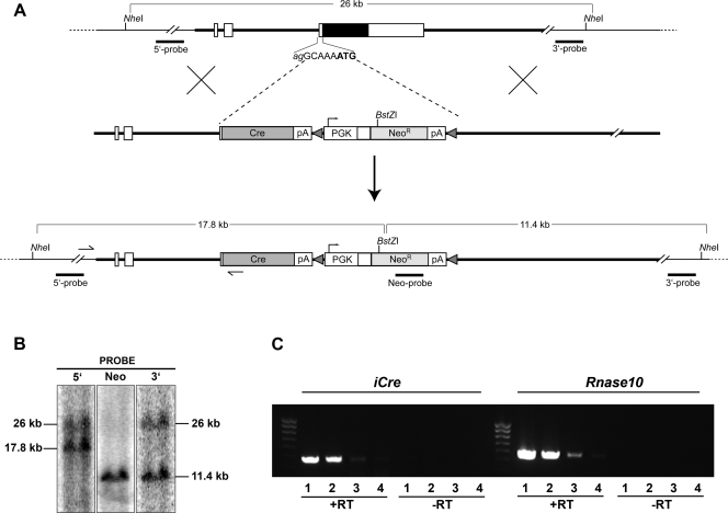
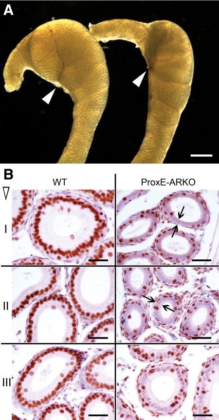
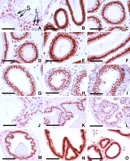
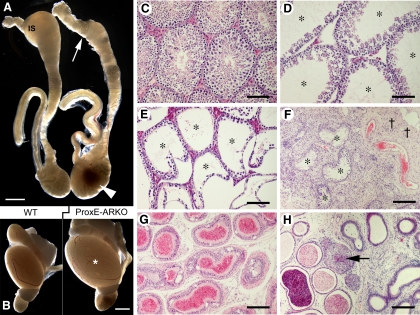
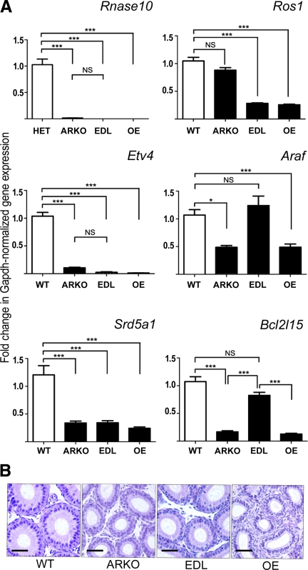
The epithelial lining of the epididymal duct expresses the androgen receptor (Ar) along its entire length and undergoes rapid and profound degeneration when androgenic support is withdrawn. However, experiments involving orchidectomy with systemic testosterone replacement, and testicular efferent duct ligation, have indicated that structural and functional integrity of the initial segment cannot be maintained by circulating androgen alone, leaving the role of androgen in this epididymal zone unclear. We addressed this question in a mouse model with intact testicular output and selective Ar inactivation in the proximal epididymis by creating double-transgenic males carrying a conditional Ar(loxP) allele and expressing Cre recombinase under the promoter of Rnase10, a gene specifically expressed in proximal epididymis. At 20-25 d of life, on the onset of Rnase10 expression, Ar became selectively inactivated in the principal cells of proximal epididymis, resulting in epithelial hypoplasia and hypotrophy. Upon the subsequent onset of spermiation, epididymal obstruction ensued, with the consequent development of spermatic granulomata, back pressure-induced atrophy of the seminiferous epithelium, orchitis, and fibrosis of the testicular parenchyma. Consistent with these findings, the mice were infertile. When the effect of Ar knockout on gene expression in the proximal epididymis was compared with that of efferent duct ligation and orchidectomy, we identified genes specifically regulated by androgen, testicular efferent fluid, and both. Our findings demonstrate that the development and function of the epididymal initial segment is critically dependent on direct androgen regulation. The phenotype of the produced knockout mouse provides a novel model for obstructive azoospermia.


Figures

Fig. 1.
Fig. 1.
Targeted insertion of iCre into the Rnase10 locus. A, Diagrammatic representation of the targeting strategy showing the wild-type Rnase10 locus (top panel), the targeting vector (middle panel), and the modified allele (bottom panel). The gene comprises two exons with alternative transcription start sites; the coding sequence shown as a filled box resides entirely in the second exon. The targeting vector introduces the iCre-NeoR cassette instead of the first eight nucleotides of exon 2, which include the Rnase10 translation initiation site (shown in bold font); the endogenous splice acceptor is retained, whereas the iCre open reading frame is headed by a strong Kozak consensus sequence. The positive selection cassette, PGK-NeoR, is flanked by minimal FRT sites (shown as shaded triangles) enabling its subsequent removal. Both iCre and NeoR are appended with sequences containing a polyadenylation signal (pA). Bold lines represent stretches of homology; arrows indicate annealing sites for the PCR primers used for initial screening of geneticin-resistant embryonic stem (ES) cell clones. Also shown are diagnostic restriction sites and genomic probes for Southern blot analysis. B, Southern blot analysis of the heterozygous ES cell clone used for blastocyst injection. Genomic DNA digested with NheI and BstZI was successively hybridized to radiolabeled probes as shown in A. C, RT-PCR analysis of iCre and Rnase10 transcripts in the proximal epididymis of the Rnase10Cre/+ male. Total RNA was isolated individually from segments I, II, III, and IV (lanes 1–4) and subjected to RT-PCR. iCre transcript was identified with the pair of specific primers detailed in Materials and Methods; oligonucleotides used for the detection of Rnase10 mRNA were 5′-GTATGGAGAGCAGTTTCTGG-3′ and 5′-CCGTTACAGACAGCTTTGAC-3′. RNA samples were not treated with deoxyribonuclease I; therefore, control reactions were performed with the omission of reverse transcriptase (−RT).
Fig. 2.
Fig. 2.
Morphological appearance of the proximal epididymis in ProxE-ARKO mice. A, Dorsal view of the proximal epididymides of WT (left) and ProxE-ARKO (right) littermates at 30 dpp; the arrowheads indicate points of entry of the superior epididymal artery demarcating the IS distally; hypoplasia of the knockout proximal epididymis is demonstrated. Bar, 1 mm. B (immunostaining with anti-AR), The proximal segments (I-III) of the epididymides of 40-d-old ProxE-ARKO (right) and WT littermates (left) are shown; arrows indicate the reduced volume of principal cells devoid of AR protein immunoreactivity in the knockout specimens. Bar, 50 μm.
Fig. 3.
Fig. 3.
Histological appearance and immunohistochemical detection of AR protein in reproductive organs of ProxE-ARKO mice. At 40 dpp: A, testis (S, Sertoli cells; L, Leydig cells); B, efferent ducts; C–I, epididymis segments IV-X; at 60 dpp: J, vas deferens; K, seminal vesicle; L, coagulating gland; M, ampullary gland; N, ventral prostate; O, dorsal prostate. Bar, 50 μm (counterstaining with hematoxylin). Immunodetection of AR in WT male reproductive tract is shown in Supplemental Fig. 4.
Fig. 4.
Fig. 4.
Consequences of Ar ablation in the proximal epididymis. A, ProxE-ARKO epididymis on the right shows accumulation of inspissated spermatozoa in the hypoplastic proximal part (arrow) and a spermatic granuloma in the caudal region (arrowhead); a wild-type control epididymis is shown on the left. Bar, 2 mm. B, Testicular outflow obstruction results in dilatation of seminiferous tubules and increased testis size (asterisk) in the ProxE-ARKO male (right); wild-type control is shown on the left. Bar, 3 mm. C–H, Hematoxylin and eosin. C, Control testis. Bar, 100 μm. D and E, Progressive dilated atrophy of germinal epithelium; lumen of seminiferous tubules is indicated with asterisks. Bar, 100 μm. F, Orchitis and interstitial fibrosis (crosses) in ProxE-ARKO testis; asterisks indicate seminiferous tubules, some containing intraepithelial cysts. Bar, 200 μm. G, Degenerative changes in cauda epididymidis. Bar 150 μm. H, Focus of granulomatous inflammation in distal caput epididymidis; arrow indicates spermatozoa in the center of spermatic granuloma. Bar, 150 μm.
Fig. 5.
Fig. 5.
A, Expression of Rnase10, Etv4, Srd5a1, Ros1, Araf, and Bcl2l15 in the IS of intact WT, ProxE-ARKO (ARKO), and EDL and OE wild-type mice. Each bar is the mean ± sem of measurements from three animals. Statistical significances: ***, P < 0.001; *, P < 0.05. NS, Not significant. The micrographs at the bottom (B) show representative views of proximal epididymis of the four experimental groups. Bar, 30 μm.

Similar articles

Cited by

References

    1. Robaire B, Hinton BT, Orgebin-Crist MC. 2006. The epididymis. In: Neill JD. ed. Knobil and Neilly's physiology of reproduction. Vol 1 San Diego: Elsevier Academic Press; 1071–1148
    1. Fawcett DW, Hoffer AP. 1979. Failure of exogenous androgen to prevent regression of the initial segments of the rat epididymis after efferent duct ligation or orchidectomy. Biol Reprod 20:162–181 - PubMed
    1. Hamzeh M, Robaire B. 2009. Effect of testosterone on epithelial cell proliferation in the regressed rat epididymis. J Androl 30:200–212 - PubMed
    1. Zhou Q, Nie R, Prins GS, Saunders PT, Katzenellenbogen BS, Hess RA. 2002. Localization of androgen and estrogen receptors in adult male mouse reproductive tract. J Androl 23:870–881 - PubMed
    1. De Gendt K, Swinnen JV, Saunders PT, Schoonjans L, Dewerchin M, Devos A, Tan K, Atanassova N, Claessens F, Lécureuil C, Heyns W, Carmeliet P, Guillou F, Sharpe RM, Verhoeven G. 2004. A Sertoli cell-selective knockout of the androgen receptor causes spermatogenic arrest in meiosis. Proc Natl Acad Sci USA 101:1327–1332 - PMC - PubMed

Publication types