. 2011 Jan;152(1):303-12.
doi: 10.1210/en.2010-0749. Epub 2010 Nov 17.

Gene expression profiling reveals Cyp26b1 to be an activin regulated gene involved in ovarian granulosa cell proliferation

Affiliations

Gene expression profiling reveals Cyp26b1 to be an activin regulated gene involved in ovarian granulosa cell proliferation

Jingjing L Kipp et al. Endocrinology. 2011 Jan.

Abstract

Activin, a member of the TGF-β superfamily, is an important modulator of FSH synthesis and secretion and is involved in reproductive dysfunctions and cancers. It also regulates ovarian follicle development. To understand the mechanisms and pathways by which activin regulates follicle function, we performed a microarray study and identified 240 activin regulated genes in mouse granulosa cells. The gene most strongly inhibited by activin was Cyp26b1, which encodes a P450 cytochrome enzyme that degrades retinoic acid (RA). Cyp26b1 has been shown to play an important role in male germ cell meiosis, but its expression is largely lost in the ovary around embryonic d 12.5. This study demonstrated that Cyp26b1 mRNA was expressed in granulosa cells of follicles at all postnatal developmental stages. A striking inverse spatial and temporal correlation between Cyp26b1 and activin-βA mRNA expression was observed. Cyp26b1 expression was also elevated in a transgenic mouse model that has decreased activin expression. The Cyp26 inhibitor R115866 stimulated the proliferation of primary cultured mouse granulosa cells, and a similar effect was observed with RA and activin. A pan-RA receptor inhibitor, AGN194310, abolished the stimulatory effect of either RA or activin on granulosa cell proliferation, indicating an involvement of RA receptor-mediated signaling. Overall, this study provides new insights into the mechanisms of activin action in the ovary. We conclude that Cyp26b1 is expressed in the postnatal mouse ovary, regulated by activin, and involved in the control of granulosa cell proliferation.


Figures

Figure 1
Figure 1
A heat map overview of all the genes that are significantly regulated by activin A as identified by Illumina BeadArray (n = 4). Red, Up-regulation; green, down-regulation. Act, Activin A; Fst, follistatin.
Figure 2
Figure 2
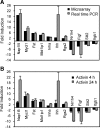
Validation of microarray results and time-course study of activin effect on selected genes. A, Validation of select activin up- and down-regulated genes obtained from microarray results with real-time RT-PCR (P < 0.05 for all; microarray, n = 4; real-time RT-PCR, n = 3–5). B, Time-course study of activin effect on selected genes. The same data for 24 h from A are plotted here as part of the time-course comparison. Primary cultured granulosa cells were treated with activin A or PBS (control) for 4 or 24 h, and mRNA levels were measured with real-time RT-PCR (P < 0.05 for all, n = 3–5).
Figure 3
Figure 3
Localization and relative expression levels of Cyp26b1 in the mouse ovary. A–D, In situ hybridization of Cyp26b1 in postnatal d 1 (A), 6 (B), 10 (C), and 20 (D) ovaries. Insert (A), Enlarged picture of two primordial follicles. E, top panel, Western blot showing the Cyp26b1 protein levels in d 1 testes and ovaries. Actin was detected as a loading control. Bottom panel, Comparison of Cyp26b1 mRNA levels in d 1 ovaries and testes as measured by real-time PCR. **, P < 0.005 (n = 4). F, Comparison of Cyp26b1 mRNA levels with inhibin-α (Inha), follistatin (Fst), ERβ (Esr2), and Cyp19a1 in d 22 mouse granulosa cells as measured by real-time PCR. Consistent amplification efficiency was observed for the primers for Inha, Fst, and Esr2 in the d 22 granulosa cell samples and for Cyp19a1 in adult ovaries (efficiency = 100 ± 3%, R2 = 0.990–0.999). Different letters indicate statistically significant differences, according to ANOVA followed by Tukey’s test (P < 0.05, n = 3–8).
Figure 4
Figure 4
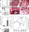
Activin regulation of Cyp26b1 gene expression and the inverse correlation of Cyp26b1 and activin βA mRNA expression. A, Effects of activin A (Act), activin A+follistatin (Act+Fst), follistatin (Fst) alone, and TGF-β1 on Cyp26b1 mRNA levels in primary cultured granulosa cells (P < 0.05 for all, n = 3). B, Effect of activin A (Act) on Cyp26b1 protein levels. Actin was detected as a loading control. C, Comparison of Cyp26b1 mRNA levels in MT-α inhibin transgenic (MT-α Inh) mouse ovaries with those in the NLM ovaries. ***, P < 0.0001 (n = 7). D, Western blot pictures showing Cyp26b1 protein levels in NLM and MT-α Inh mouse ovaries. Actin was detected as a loading control. E–H, In situ hybridization of Cyp26b1 (E and G) and activin-βA (F and H) in postnatal d 20 ovaries. E and F show lower magnification; G and H show higher magnification. Complementary expression patterns of Cyp26b1 and activin-βA mRNA within one follicle is indicated by open arrows. GC, Granulosa cells. Oo, oocytes. I, Comparison of mRNA levels of Cyp26b1, activin-βA (Inhba), and activin-βB (Inhbb) in developing ovaries as measured by real-time PCR. Different letters indicate statistically significant differences, according to ANOVA followed by Tukey’s test (lowercase letters for Cyp26b1; uppercase letters for activin βA). P < 0.05 (n = 3–5).
Figure 5
Figure 5
Effects of RA (A) and the Cyp26 inhibitor, R115866 (B) on proliferation of primary cultured mouse granulosa cells. *, P < 0.05; ***, P < 0.005 (n = 3–7).
Figure 6
Figure 6
Effects of the pan-RAR inhibitor, AGN193109, on RA- (A) or activin A (B)-induced proliferation of primary cultured mouse granulosa cells and lack of effect of AGN193109 on select activin target genes (C). Con, Control; Act, activin A. *, P < 0.05 (n = 3–5).

Similar articles

Cited by

References

    1. McGee EA, Hsueh AJ 2000 Initial and cyclic recruitment of ovarian follicles. Endocr Rev 21:200–214 - PubMed
    1. Edson MA, Nagaraja AK, Matzuk MM 2009 The mammalian ovary from genesis to revelation. Endocr Rev 30:624–712 - PMC - PubMed
    1. Epifano O, Dean J 2002 Genetic control of early folliculogenesis in mice. Trends Endocrinol Metab 13:169–173 - PubMed
    1. Barnett KR, Schilling C, Greenfeld CR, Tomic D, Flaws JA 2006 Ovarian follicle development and transgenic mouse models. Hum Reprod Update 12:537–555 - PubMed
    1. Richards JS, Pangas SA 2010 The ovary: basic biology and clinical implications. J Clin Invest 120:963–972 - PMC - PubMed

Publication types

MeSH terms