. 2011 Feb;85(3):1298-309.
doi: 10.1128/JVI.01957-10. Epub 2010 Nov 17.

The arginine clusters of the carboxy-terminal domain of the core protein of hepatitis B virus make pleiotropic contributions to genome replication

Affiliations

The arginine clusters of the carboxy-terminal domain of the core protein of hepatitis B virus make pleiotropic contributions to genome replication

Eric B Lewellyn et al. J Virol. 2011 Feb.

Abstract

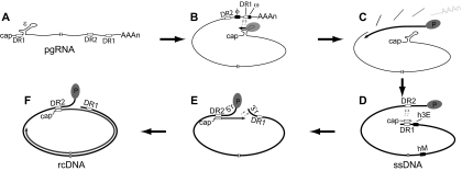
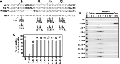
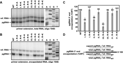
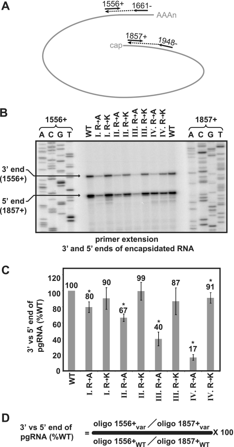
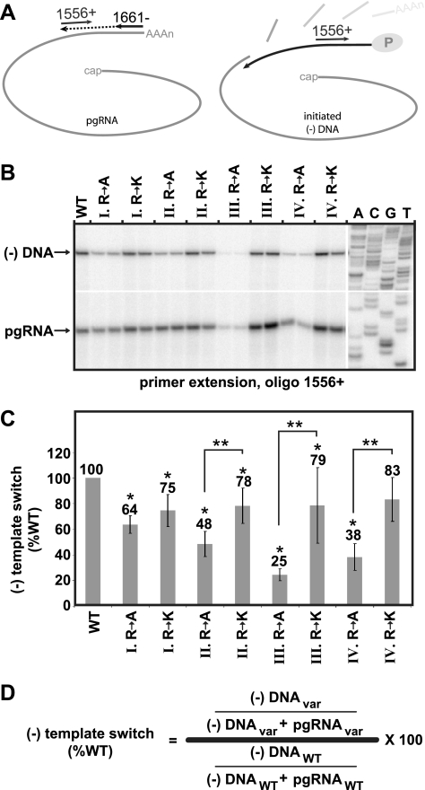
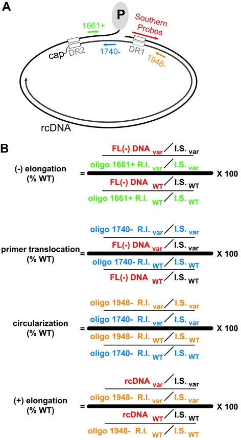
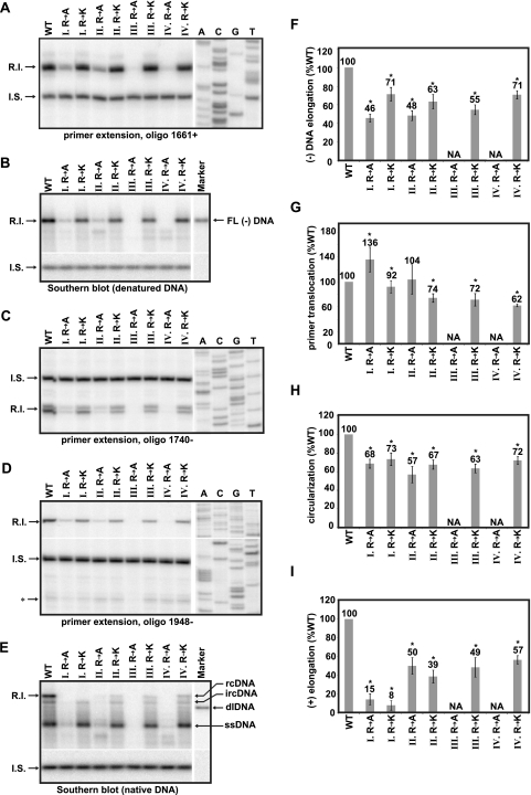
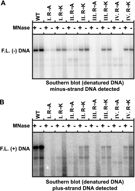
The carboxy-terminal domain (CTD) of the core protein of hepatitis B virus is not necessary for capsid assembly. However, the CTD does contribute to encapsidation of pregenomic RNA (pgRNA). The contribution of the CTD to DNA synthesis is less clear. This is the case because some mutations within the CTD increase the proportion of spliced RNA to pgRNA that are encapsidated and reverse transcribed. The CTD contains four clusters of consecutive arginine residues. The contributions of the individual arginine clusters to genome replication are unknown. We analyzed core protein variants in which the individual arginine clusters were substituted with either alanine or lysine residues. We developed assays to analyze these variants at specific steps throughout genome replication. We used a replication template that was not spliced in order to study the replication of only pgRNA. We found that alanine substitutions caused defects at both early and late steps in genome replication. Lysine substitutions also caused defects, but primarily during later steps. These findings demonstrate that the CTD contributes to DNA synthesis pleiotropically and that preserving the charge within the CTD is not sufficient to preserve function.


Figures

FIG. 1.
FIG. 1.
DNA synthesis for HBV. (A) The pgRNA is greater than the genome length and has 5′ and 3′ copies of the 11-nt direct repeat sequence DR1 and the stem-loop ɛ. The relative position of the other direct repeat sequence, DR2, is also indicated. pgRNA is translated to make the core protein and P protein. Dimers of the core protein assemble into capsids. P protein associates with ɛ and the combination of the two comprises the encapsidation signal. (B) (−)DNA template switch. Minus-strand DNA synthesis initiates within the bulge of ɛ. Tyrosine 63 of P protein is the primer for the first 3 to 4 nt and the bulge of ɛ is the template. The minus-strand then switches template to an acceptor site within the 3′ copy of DR1. Base pairing between nucleotides in the upper stem of ɛ and cis-acting sequences φ and ω contribute to these steps. (C) (−)DNA elongation. Minus-strand DNA elongation and concomitant RNase H degradation of the pgRNA are catalyzed by the P protein, resulting in a full-length, single-stranded DNA. The 5′-most end of the pgRNA remains undigested. (D) Primer translocation. The pgRNA remnant undergoes a template switch from DR1 to DR2 and primes plus-strand DNA synthesis from DR2. The process cannot proceed without cis-acting sequences h3E and hM, which base pair with one another. Several other cis-acting sequences make additional contributions to the process (41). (E) Circularization. After synthesizing to the 5′ end of the minus-strand DNA template, the 3′ end of the plus-strand switches templates to use the 3′ end of minus-strand DNA. Like primer translocation, this step requires base pairing between cis-acting sequences h3E and hM (39). (F) (+) Elongation. Once annealed to 3′r, the plus-strand synthesis resumes, ultimately yielding rcDNA.
FIG. 2.
FIG. 2.
Mutations within the CTD have little to no impact on capsid assembly. (A) Alignment of the sequence of CTD of HBV, woolly monkey hepatitis B virus (WMHBV), woodchuck hepatitis B (WHV), and ground squirrel hepatitis B virus (GSHV). Boldface residues indicate differences from the HBV sequence. The sequences of WMHBV, GSHBV, and WHV are from GenBank accession numbers AAO74859.1, K02715.1, and AY628095.1, respectively. The positions of four clusters of arginines—I, II, III, and IV—are also shown. Variants of the HBV core protein are indicated below. Arginines were replaced with either alanines or lysines in each variant. (B) Western blot analysis of velocity sedimentation of cytoplasmic lysates from cells transfected with each of the core variants. Core protein was detected, and a molecular weight marker with bands corresponding to ∼15 and ∼25 kDa is shown in the left lane of each blot (MWM). (C) Quantitation of assembly. Fractions 9 to 13 are considered to be unassembled or improperly assembled core based on comparison to Y132A, a variant that is known to not form capsids (10). For each variant, the proportion of core in fractions 1 to 8 (assembled) is shown as a percent total core protein. Error bars represent ± the 95% confidence interval. Each sample was analyzed a minimum of four times. An asterisk (*) indicates a significant difference from the WT reference (P < 0.05).
FIG. 3.
FIG. 3.
Encapsidation of pgRNA. Primer extension using oligo 1948− extended with avian myeloblastosis virus (AMV) reverse transcriptase (RT) was used to measure the level of 5′ ends of encapsidated and reference RNAs. (A and B) Representative gels used to measure the encapsidation efficiency of total and encapsidated pgRNA, respectively. In each gel, the positions of the pgRNA and reference RNA (ref. RNA) are indicated. A sequencing ladder is provided for reference. (C) Histogram of the efficiency of encapsidation of the 5′ end of pgRNA, with standard deviations of normalized values represented by error bars. (D) Formula used to calculate encapsidation. An asterisk (*) indicates a significant difference from the WT reference (P < 0.05).
FIG. 4.
FIG. 4.
Ratio of 3′ to 5′ ends of encapsidated pgRNA. Sequential RNA and DNA primer extension was used to determine the ratio of encapsidated 3′ to 5′ ends of pgRNA. Unlabeled oligos 1948− and 1661− were used to generate cDNAs corresponding to the 5′ and 3′ ends. Radiolabeled oligos 1857+ and 1556+ were subsequently used to detect those cDNAs. (A) A diagram showing this strategy is shown. (B) Representative gel used to determine the ratio of 3′ to 5′ ends. The position of the 3′ and 5′ ends are indicated. Sequencing ladders corresponding to both labeled primers are shown on either side of the gel. (C) A histogram shows the ratio of each variant relative to the wild-type reference. (D) Formula used to calculate the ratio of 3′ to 5′ ends. An asterisk (*) indicates a significant difference from the WT reference (P < 0.05).
FIG. 5.
FIG. 5.
Efficiency of minus-strand DNA template switch. (A) The positions of two oligonucleotide primers are shown. Primer 1661− was used to generate a cDNA corresponding to the 3′ end of pgRNA. Radiolabeled oligonucleotide 1556+ was used to detect both the cDNA and minus-strand DNA that has undergone the template switch to DR1. (B) Representative dual primer extension gel with positions of the cDNA corresponding to the pgRNA and minus-strand DNA indicated. A sequencing ladder is shown as a reference. (C) Histogram showing the relative efficiency of (−)DNA template switch of each of the variants as calculated by using the equation shown in panel D. An asterisk (*) indicates a significant difference from the WT reference (P < 0.05). Double asterisks (**) indicate a significant difference between bracketed variants (P < 0.05).
FIG. 6.
FIG. 6.
Calculation of efficiency of different steps or stages of DNA synthesis. (A) Position of all oligonucleotide primers and Southern blot hybridization probes used to detect DNA at various stages. Primer extension primers 1661+ (green), 1740+ (blue), and 1948− (orange) were used to detect (−)DNA initiated from DR1, DR2-primed (+)DNA, and circularized (+)DNA, respectively. Southern blotting was used to detect full-length (−)DNA and rcDNA. (B) Equations for calculation of (−) elongation, primer translocation, circularization, (+) elongation. FL (−)DNA is the amount of full-length minus-strand DNA, as detected by Southern blotting (red). Oligo 1661+ R.I. is the amount of minus-strand DNA elongating from DR1, as determined by primer extension with oligon 1661+ (green). Oligo 1740− R.I. is the amount of plus-strand DNA that is initiated at DR2, as detected by primer extension with oligo 1740− (blue). Oligo 1948− R.I. is the amount of plus-strand DNA that is initiated at DR2 and circularized, as detected by primer extension with oligo 1948− (orange). In all cases, a common internal standard DNA (I.S.) was used to normalize and compare the level of viral DNA detected by the different assays and methods. The probes to detect the Southern blot I.S. are specific for a sequence that is not within the HBV genome. A subscript “var” indicates the calculation for a variant. A subscript “WT” indicates the calculation for the wild-type reference. The color of each variable is matched to the probe or primer used to detect that replicative intermediate.
FIG. 7.
FIG. 7.
Analysis of DNA synthesis efficiency during functionally distinct stages. For all gel images, the positions of the replicative intermediate (R.I.) and internal standard (I.S.) bands that were measured are indicated. (A) Primer extension with oligo 1661+ to detect minus-strand DNA elongated from DR1. (B) Southern blot with heat-denatured DNA to detect full-length minus-strand DNA. (C) Primer extension with oligo 1740− to detect plus-strand DNAs initiated from DR2. (D) Primer extension with oligo 1948− to detect circularized, DR2-initiated plus-strand DNAs. The position of in situ-primed plus-strand DNAs is indicated (*). (E) Southern blot with native DNA. The positions of ssDNA, dlDNA, ircDNA, and full-length rcDNA are indicated. (F thru I). Histograms showing relative efficiency of (−)DNA elongation, primer translocation, circularization, and (+)DNA elongation, as calculated by the equations in Fig. 6. Samples III R to A and IV R to A were not analyzed (NA) because DNA levels were below the limits of quantitative detection due to accumulation of defects at earlier steps in replication. An asterisk (*) indicates a statistically significant difference from the WT reference (P < 0.05).
FIG. 8.
FIG. 8.
Capsid mutations do not increase susceptibility of DNA to MNase. HBV DNA was isolated using either MNase or MNase omitted. After isolation, all samples were treated with RNase A and DpnI to digest transfected plasmid DNA and RNA. Subsequent Southern blot analysis on heat-denatured DNA was conducted. Southern blots were probed with minus-strand-specific probes (oligos 1816+, 1833+, 1857+, and 1876+) (A) or a plus-strand-specific riboprobe that spans the full genome length (B).

Similar articles

Cited by

References

    1. Abraham, T. M., E. B. Lewellyn, K. M. Haines, and D. D. Loeb. 2008. Characterization of the contribution of spliced RNAs of hepatitis B virus to DNA synthesis in transfected cultures of Huh7 and HepG2 cells. Virology 379:30-37. - PMC - PubMed
    1. Abraham, T. M., and D. D. Loeb. 2006. Base pairing between the 5′ half of epsilon and a cis-acting sequence, phi, makes a contribution to the synthesis of minus-strand DNA for human hepatitis B virus. J. Virol. 80:4380-4387. - PMC - PubMed
    1. Abraham, T. M., and D. D. Loeb. 2007. The topology of hepatitis B virus pregenomic RNA promotes its replication. J. Virol. 81:11577-11584. - PMC - PubMed
    1. Auxilien, S., G. Keith, S. F. Le Grice, and J. L. Darlix. 1999. Role of posttranscriptional modifications of primer tRNALys,3 in the fidelity and efficacy of plus strand DNA transfer during HIV-1 reverse transcription. J. Biol. Chem. 274:4412-4420. - PubMed
    1. Bartenschlager, R., M. Junker-Niepmann, and H. Schaller. 1990. The P gene product of hepatitis B virus is required as a structural component for genomic RNA encapsidation. J. Virol. 64:5324-5332. - PMC - PubMed

Publication types

LinkOut - more resources