. 2011 Feb;85(3):1224-36.
doi: 10.1128/JVI.01635-10. Epub 2010 Nov 17.

RIG-I-mediated antiviral signaling is inhibited in HIV-1 infection by a protease-mediated sequestration of RIG-I

Affiliations

RIG-I-mediated antiviral signaling is inhibited in HIV-1 infection by a protease-mediated sequestration of RIG-I

Mayra Solis et al. J Virol. 2011 Feb.

Abstract

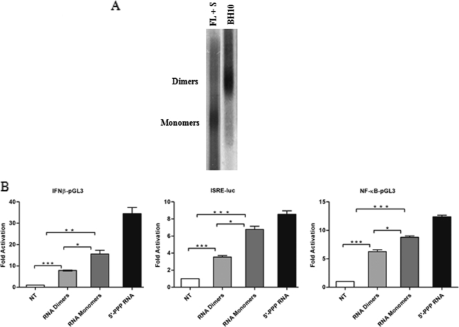
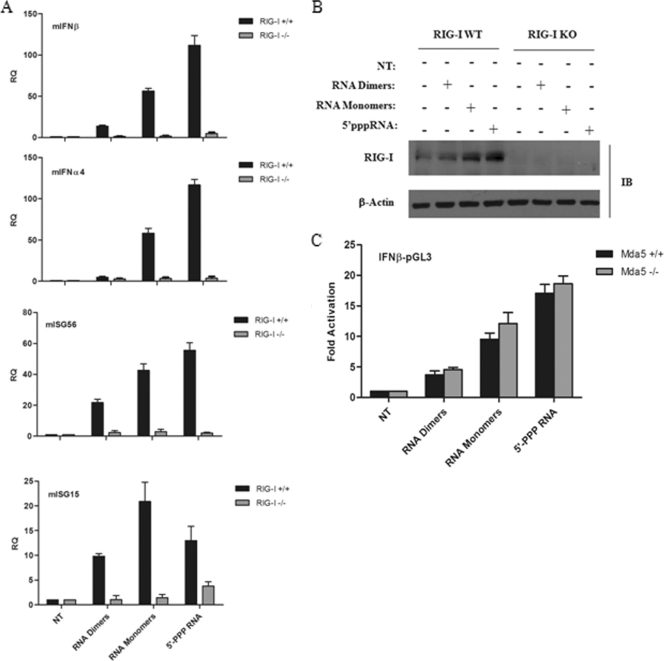
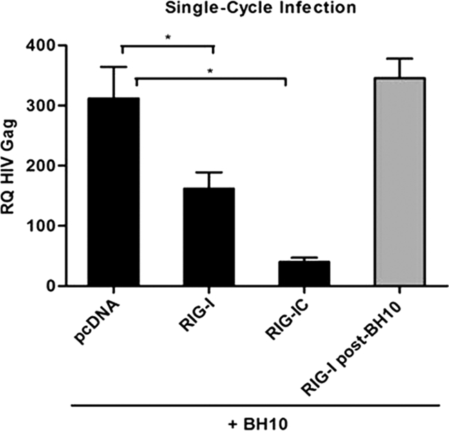
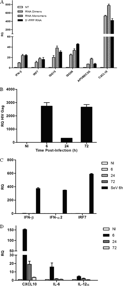
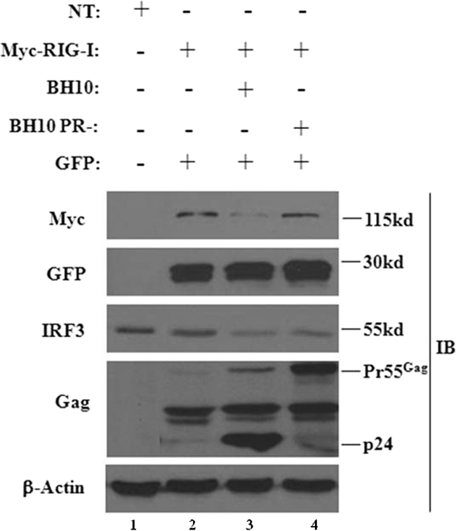
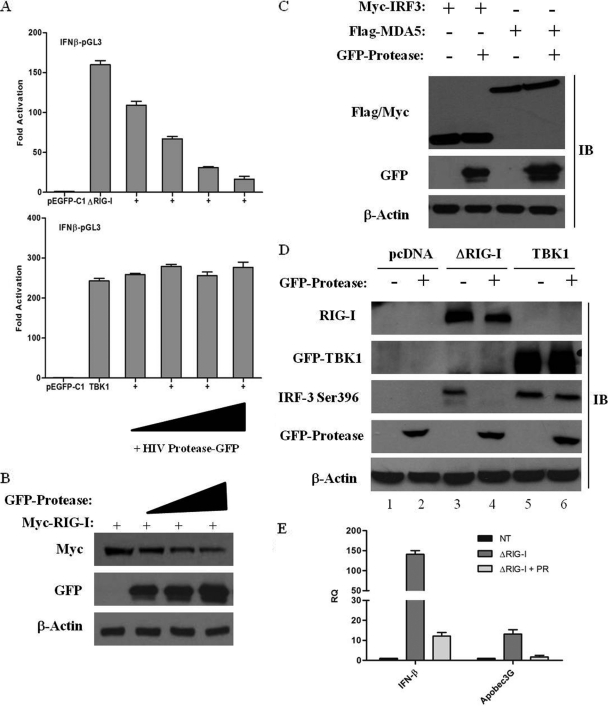
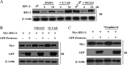
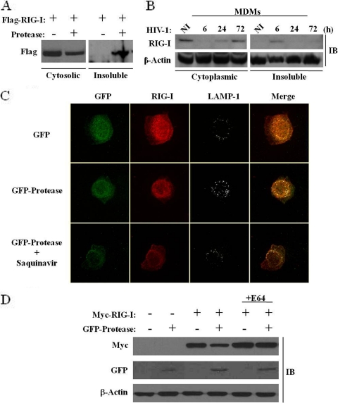
The rapid induction of type I interferon (IFN) is essential for establishing innate antiviral responses. During infection, cytoplasmic viral RNA is sensed by two DExD/H box RNA helicases, RIG-I and MDA5, ultimately driving IFN production. Here, we demonstrate that purified genomic RNA from HIV-1 induces a RIG-I-dependent type I IFN response. Both the dimeric and monomeric forms of HIV-1 were sensed by RIG-I, but not MDA5, with monomeric RNA, usually found in defective HIV-1 particles, acting as a better inducer of IFN than dimeric RNA. However, despite the presence of HIV-1 RNA in the de novo infection of monocyte-derived macrophages, HIV-1 replication did not lead to a substantial induction of IFN signaling. We demonstrate the existence of an evasion mechanism based on the inhibition of the RIG-I sensor through the action of the HIV-1 protease (PR). Indeed, the ectopic expression of PR resulted in the inhibition of IFN regulatory factor 3 (IRF-3) phosphorylation and decreased expression of IFN and interferon-stimulated genes. A downregulation of cytoplasmic RIG-I levels occurred in cells undergoing a single-cycle infection with wild-type provirus BH10 but not in cells transfected with a protease-deficient provirus, BH10-PR(-). Cellular fractionation and confocal microscopy studies revealed that RIG-I translocated from the cytosol to an insoluble fraction during the de novo HIV-1 infection of monocyte-derived macrophages, in the presence of PR. The loss of cytoplasmic RIG-I was prevented by the lysosomal inhibitor E64, suggesting that PR targets RIG-I to the lysosomes. This study reveals a novel PR-dependent mechanism employed by HIV-1 to counteract the early IFN response to viral RNA in infected cells.


Figures

FIG. 1.
FIG. 1.
HIV-1 genomic RNA induces IFN signaling. (A) HIV-1 viral RNA dimers were obtained from WT BH10 virions, whereas HIV-1 RNA monomers were isolated from BH10 virions grown in the presence of the protease inhibitor saquinavir (S) (1 μM) for 48 h. Also, a joint mutation in the amino-terminal finger and linker region of the nucleocapsid was included in these virions. Genomic RNAs extracted from the respective virions were electrophoresed on a nondenaturing 1% (wt/vol) agarose gel and analyzed by Northern blotting. (B) Activity of type I IFN promoters by HIV-1 RNA. HeLa cells were transfected with the respective reporter plasmids for IFNB-pGL3, ISRE-Luc, and NF-κB-pGL3. At 24 h posttransfection, HIV-1 gRNA dimers, monomers, or control 5′-PPP RNA was transfected. Luciferase activity was analyzed at 24 h posttransfection. Values are as means ± SEM. Statistical significance was evaluated by using P values of <0.05 (*), <0.01 (**), and <0.001 (***). NT, not treated.
FIG. 2.
FIG. 2.
RIG-I recognizes both dimeric and monomeric forms of full-length HIV-1 viral RNA. (A) RIG-I+/+ and RIG-I−/− mouse embryo fibroblasts (MEFs) were transfected with HIV-1 gRNA dimers or monomers along with control 5′-PPP RNA, respectively. Total RNA was isolated at 24 h posttransfection and subjected to real-time PCR analysis for the quantification of type I IFN gene expression, such as mIFN-β, mIFN-α4, mISG15, mISG56, and mβ-actin. Results are presented as relative quantification (RQs). Values are means ± SEM. (B) Whole-cell extracts from RIG-I+/+ (WT) and RIG-I−/− (KO) MEFs transfected with HIV-1 gRNA dimers or monomers along with control 5′-PPP RNA, respectively, were subjected to Western blot analysis and probed with anti-RIG-I and anti-β-actin antibodies. (C) The IFNB-pGL3 promoter construct was transfected into MDA5+/+ and MDA5−/− MEFs. Subsequently, HIV-1 gRNA dimers, monomers, or control 5′-PPP RNA was transfected. IFNB promoter activity was analyzed at 24 h posttransfection. Values represent means ± SEM. IB, immunoblot.
FIG. 3.
FIG. 3.
RIG-I overexpression inhibits HIV-1 replication in single-cycle infections. Total RNA was extracted from HEK293 cells 24 h after cotransfection of pcDNA3, Myc-RIG-I, and Myc-RIG-I C along with the BH10 proviral clone. BH10 transfection prior to Myc-RIG-I transfection is shown by the gray bar. cDNA samples were subjected to real-time PCR analysis with primers specific for the HIV-1 Gag gene or β-actin. HIV-1 replication is represented as an RQ based on the relative expression of the HIV-1 Gag gene versus β-actin as a reference gene. Values are means ± SEM. Statistical relevance was evaluated using a P value of <0.05 (*).
FIG. 4.
FIG. 4.
HIV-1 viral RNA induces type I IFNs and interferon-stimulated genes in macrophages. (A) PMA-differentiated THP-1 cells were transfected with HIV-1 RNA dimers, RNA monomers, or RNA bearing 5′-triphosphate for 24 h, respectively. Total RNA was isolated at 24 h posttransfection and subjected to real-time PCR analysis for the quantification of IFN-stimulated gene expression, such as IFN-β, IRF-7, ISG15, ISG56, APOBEC3G, and β-actin. (B) Kinetics of HIV-1 replication in monocyte-derived macrophages (MDMs). MDMs were infected de novo (or not) with HIV-1 at an MOI of 1 for 6, 24, and 72 h. Total RNA was isolated at the indicated times and subjected to real-time PCR analysis with primers specific for the HIV-1 Gag gene or β-actin. (C and D) De novo HIV-1 infection of MDMs does not induce a type I IFN response but triggers a proinflammatory response. MDMs were infected de novo (or not) with HIV-1 at an MOI of 1 for 6, 24, and 72 h. MDMs infected with Sendai virus (40 hemagglutinating units/ml) for 6 h were used as a control for the induction of type I IFNs. Expression levels of IFN-β, IFN-α2, IRF-7, CXCL10, IL-12α, IL-6, and β-actin were analyzed by real-time PCR. Results are presented as RQs. Values are means ± SEM. NI, not infected.
FIG. 5.
FIG. 5.
An HIV-1 protease-deficient provirus restores the expression of RIG-I. HEK293 cells were transfected with a plasmid expressing Myc-RIG-I. At 24 h posttransfection cells were transfected with either the WT BH10 proviral clone or the BH10-protease-defective proviral clone. A GFP-expressing plasmid alone was used as an internal control. Whole-cell extracts were subjected to Western blot analysis using anti-Myc, anti-GFP, anti-IRF-3, anti-p24 Gag, and anti-β-actin antibodies. The 55-kDa band represents the immature Gag polyprotein, whereas the 24-kDa band represents the mature Gag protein.
FIG. 6.
FIG. 6.
HIV-1 PR targets RIG-I and interferes with IFN-β activation. (A) HIV-1 PR downregulates the IFNB promoter. HEK293 cells were transfected with an IFNB-pGL3 reporter plasmid, the pEGFP-C1 vector, or expression plasmids encoding ΔRIG-I and TBK1 along with increasing amounts (50 ng, 100 ng, 200 ng, and 500 ng) of the GFP-protease expression construct. IFNB promoter activity was measured at 24 h posttransfection. Values represent means ± SEM. (B) RIG-I is downregulated in the presence of HIV-I PR. HEK293 cells were cotransfected with expression vectors for Myc-RIG-I (2 μg) and increasing amounts (2 μg, 5 μg, and 10 μg) of GFP-protease as indicated. Cell lysates were subjected to Western blot analysis, and expression levels of RIG-I, protease, and β-actin were analyzed by immunoblotting with antibodies against Myc, GFP, and β-actin, respectively. (C) HIV-1 PR specifically targets RIG-I. HEK293 cells were cotransfected with plasmids expressing Myc-IRF-3, Flag-MDA5, and GFP-protease. Cell lysates were subsequently subjected to immunoblotting with anti-Myc, anti-Flag, anti-GFP, and anti-β-actin antibodies. (D) HIV-1 PR inhibits ΔRIG-I-mediated activation of IRF-3. HEK293 cells were transfected with 500 ng of Myc-IRF-3, 1 μg Myc-ΔRIG-I or GFP-TBK1, and 1 μg of GFP-protease expression plasmids as indicated. Whole-cell extracts were analyzed by immunoblotting for IRF-3 pSer-396, RIG-I, GFP-TBK1, GFP-protease, and β-actin. (E) HIV-1 PR downregulates IFN-stimulated gene expression. HEK293 cells were transfected with Myc-ΔRIG-I along with the GFP-protease expression plasmid. cDNA samples were subjected to real-time PCR analysis with primers specific for IFN-β, APOBEC3G, and β-actin. Results are presented as RQs. Values are means ± SEM.
FIG. 7.
FIG. 7.
HIV-1 PR downregulates RIG-I protein expression through a proteasome- and caspase-independent mechanism. (A) RIG-I downregulation is proteasome and caspase independent. THP-1 cells were infected de novo with HIV-1 at an MOI of 1 for 6, 24, and 72 h. Cells were incubated with either dimethyl sulfoxide (DMSO), 5 μM MG132 (proteasomal inhibitor), or 50 μM Z-VAD (pancaspase inhibitor), respectively. THP-1 cells infected with Sendai virus (40 hemagglutinating units/ml) for 6 h were used as a control for the induction of RIG-I expression. Whole-cell extracts were subjected to immunoblotting with anti-RIG-I and anti-β-actin antibodies. (B) RIG-I downregulation is proteasome and caspase independent. HEK293 cells were cotransfected with plasmids expressing Myc-RIG-I and GFP-protease and incubated with either 5 μM MG132 (proteasomal inhibitor) or 50 μM Z-VAD (pancaspase inhibitor). Whole-cell extracts were subjected to immunoblotting with anti-Myc, anti-GFP, and anti-β-actin antibodies. (C) HIV-1 PR-mediated downregulation of RIG-I is not dependent on the HIV-1 PR catalytic activity. HEK293 cells were transfected with Myc-RIG-I or GFP-protease expression plasmids and incubated with 5 μM saquinavir. At 24 h posttransfection, whole-cell extracts were subjected to Western blot analysis and immunoblotted for Myc-RIG-I, GFP-protease, and β-actin.
FIG. 8.
FIG. 8.
HIV-1 protease expression causes a relocalization of the cytoplasmic pool of RIG-I to the lysosomes. (A and B) HEK293 cells were cotransfected with plasmids expressing Flag-RIG-I and GFP-protease (A), and MDMs were infected de novo (or not) with HIV-1 at an MOI of 1 for 6, 24, and 72 h (B). Whole-cell extracts isolated from the cytoplasmic fraction versus the insoluble fraction were run on an SDS-PAGE gel and subsequently subjected to immunoblotting with anti-Flag, anti-RIG-I, or anti-β-actin antibodies, respectively. (C) A549 cells were transfected with plasmids expressing GFP or GFP-protease with or without saquinavir (5 μM). At 15 h posttransfection, cells were, fixed, permeabilized and immunostained for endogenous RIG-I (red) or LAMP-1 (gray). Cells were visualized by confocal microscopy. (D) A lysosomal inhibitor restores RIG-I protein expression. HEK293 cells were cotransfected with Myc-RIG-I or GFP-protease expression plasmids and treated with 10 μM E64 (lysosomal-protease inhibitor cocktail). At 24 h posttransfection, expression levels of RIG-I, protease, and β-actin were analyzed by immunoblotting with antibodies against Myc, GFP, and β-actin, respectively.

Similar articles

Cited by

References

    1. Baenziger, S., et al. 2009. Triggering TLR7 in mice induces immune activation and lymphoid system disruption, resembling HIV-mediated pathology. Blood 113:377-388. - PubMed
    1. Baum, A., R. Sachidanandam, and A. Garcia-Sastre. 2010. Preference of RIG-I for short viral RNA molecules in infected cells revealed by next-generation sequencing. Proc. Natl. Acad. Sci. U. S. A. 107:16303-16308. - PMC - PubMed
    1. Beignon, A. S., et al. 2005. Endocytosis of HIV-1 activates plasmacytoid dendritic cells via Toll-like receptor-viral RNA interactions. J. Clin. Invest. 115:3265-3275. - PMC - PubMed
    1. Blanco, R., L. Carrasco, and I. Ventoso. 2003. Cell killing by HIV-1 protease. J. Biol. Chem. 278:1086-1093. - PubMed
    1. Boasso, A., and G. M. Shearer. 2008. Chronic innate immune activation as a cause of HIV-1 immunopathogenesis. Clin. Immunol. 126:235-242. - PMC - PubMed

Publication types