. 2011 Feb;85(3):1237-45.
doi: 10.1128/JVI.01234-10. Epub 2010 Nov 17.

Identification of novel endogenous betaretroviruses which are transcribed in the bovine placenta

Affiliations

Identification of novel endogenous betaretroviruses which are transcribed in the bovine placenta

Kenji Baba et al. J Virol. 2011 Feb.

Abstract

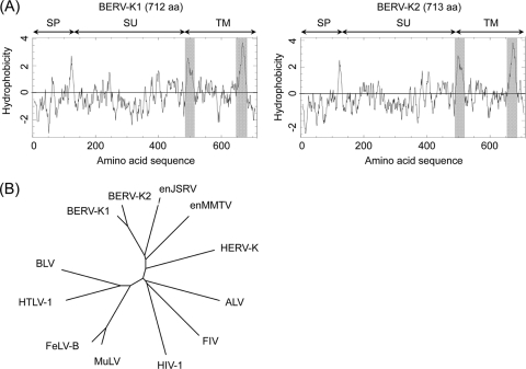
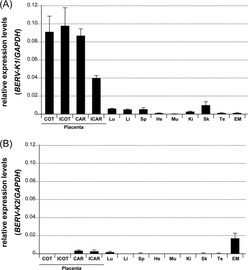
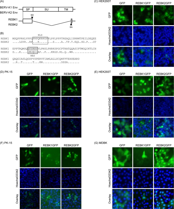
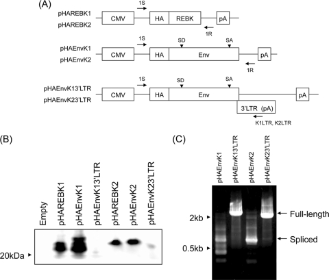
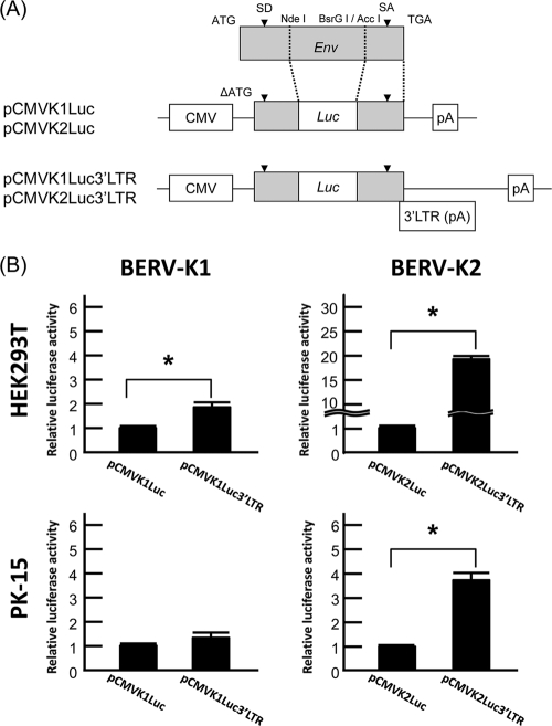
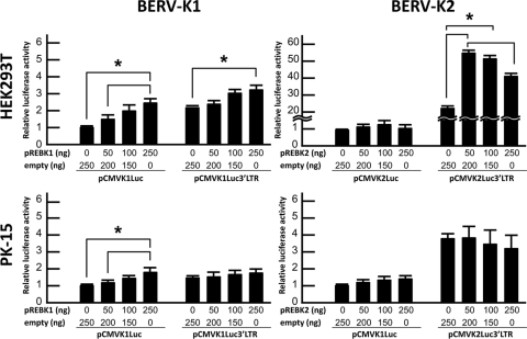
Sequences of retroviral origin occupy approximately 10% of mammalian genomes. Various infectious endogenous retroviruses (ERVs) and functional retroviral elements have been reported for several mammals but not cattle. Here, we identified two proviruses, designated bovine endogenous retrovirus K1 (BERV-K1) and BERV-K2, containing full-length envelope (env) genes in the bovine genome. Phylogenetic analysis revealed that they belong to the genus Betaretrovirus. By reverse transcription (RT)-PCR, both BERV-K1 and -K2 env mRNAs were detected in the placenta and cultured bovine trophoblast cells. Real-time RT-PCR analysis using RNAs isolated from various bovine tissues revealed that BERV-K1 env mRNA was preferentially expressed in the placenta. Moreover, we also found the expression of doubly spliced transcripts, named the REBK1 and REBK2 genes. Both the REBK1 and REBK2 proteins have motifs for a putative nuclear localization signal and a nuclear export signal. REBK1 and REBK2 fused with green fluorescent proteins were localized mainly in the nuclei when they were expressed in bovine and porcine cells. In the env and 3' long terminal repeats of BERV-K1 and -K2, we found regulatory elements responsible for the splicing and transport of viral RNAs and/or translation of the env genes. Although we have not identified the expressed Env proteins in bovine tissues, these data suggest that both BERV-K1 and BERV-K2 express Env proteins and that these proteins may have physiological functions in vivo.


Figures

FIG. 1.
FIG. 1.
Genomic structures of BERV-K1 and -K2 identified by in silico analyses. (A) Schematic representation of BERV-K1 and -K2 proviruses on bovine chromosomes 7 and 24, respectively. Arrows indicate primers used for RT-PCR analysis in B. (B) Expression of BERV-K1 and -K2 mRNAs in bovine placenta and BT-1 cells. Total RNA was extracted from the placenta and BT-1 cells and then subjected to RT-PCR analysis using the primers indicated in A. β-Actin was also amplified as an internal control.
FIG. 2.
FIG. 2.
Hydrophobicity profiles and phylogenetic analysis of Env proteins of BERV-K1 and -K2. (A) Hydrophobicity profiles of BERV-K1 and -K2 Env proteins. Gray shading indicates the fusion peptide and the transmembrane domain. (B) Phylogenetic analyses of the BERV-K1 and -K2 env genes. The phylogenetic tree of the entire TM-encoding region of env was constructed by the neighbor-joining method. enJSRV, endogenous Jaagsiekte sheep retrovirus; enMMTV, endogenous mouse mammary tumor virus; HERV-K, human endogenous retrovirus type K; ALV, avian leukemia virus; FIV, feline immunodeficiency virus; HIV-1, human immunodeficiency virus type 1; MuLV, murine leukemia virus; FeLV-B, feline leukemia virus subgroup B; HTLV-1, human T-cell leukemia virus type 1; BLV, bovine leukemia virus.
FIG. 3.
FIG. 3.
Expression of BERV-K1 and -K2 env mRNAs in normal bovine tissues. Total RNA was extracted from tissues of Holstein cows. Abbreviations: COT, cotyledon; ICOT, intercotyledon; CAR, caruncular; ICAR, intercaruncular; Lu, lung; Li, liver; Sp, spleen; He, heart; Mu, muscle; Ki, kidney; Sk, skin; Te, testis; EM, endometrium. Each value was normalized to the amount of GAPDH mRNA and is exhibited as means ± standard errors of data from three or six samples in duplicate experiments.
FIG. 4.
FIG. 4.
Identification of the REBK1 and REBK2 proteins. (A) Schematic representation of REBK1 and REBK2 transcripts. Start codons for REBK1 and REBK2 are shared with those of env. (B) Sequence alignment of the REBK1 and REBK2 proteins. Dots indicate identical amino acids. The box and the shaded box indicate NLS and NES motifs, respectively. (C to G) Subcellular localization of the REBK1 and REBK2 proteins. REBK1 and REBK2 fused with GFP (REBK1GFP and REBK2GFP, respectively) or only GFP (empty vector) was expressed in HEK293T (C and E), PK-15 (D and F), and MDBK (G) cells by transfection with expression plasmids (C and D) and infection with retroviral vectors (E, F, and G). Forty-eight hours after transfection and infection, the cells were stained with Hoechst 33342 dye and subjected to fluorescence microscopy analysis.
FIG. 5.
FIG. 5.
Splicing of BERV-K env mRNA depending on the 3′ LTR sequence. (A) Schematic representation of expression plasmids. Primers used for RT-PCR in C are indicated by arrows. (B) Immunoblot analysis for the expression of REBK1 and REBK2. HEK293T cells were transfected with the indicated expression plasmids. Forty-eight hours after transfection, cell lysates were extracted and subjected to immunoblot analysis. HA-tagged REBK1 and REBK2 were probed with a rabbit anti-HA IgG antibody and detected by an HRP-labeled mouse anti-rabbit IgG antibody. (C) Detection of full-length and spliced transcripts. Total RNAs were extracted from cells transfected with pHAEnvK1, pHAEnvK2, pHAEnvK13′LTR, and pHAEnvK23′LTR and subjected to RT-PCR analysis. Full-length env and singly spliced transcripts are indicated by arrows.
FIG. 6.
FIG. 6.
Splicing of BERV-K env mRNAs is suppressed by the 3′ LTR sequence. (A) Schematic diagrams of reporter plasmids. Plasmids contain partial sequences of BERV-K1 or BERV-K2 env without translation starting sequences (ΔATG), and the luciferase gene is inserted between the SD and SA sites of env. pCMVKLuc plasmids are Δ3′ LTR mutants of pCMVKLuc3′LTR plasmids. CMV, cytomegalovirus. (B) The relative luciferase activity of each reporter plasmid was measured in HEK293T and PK-15 cells. Values are the means ± standard errors of data from three independent experiments in triplicate. Asterisks indicate significant differences (P < 0.05).
FIG. 7.
FIG. 7.
Effects of REBKs on the splicing of BERV-K env mRNAs. Each graph indicates the relative luciferase activity of each reporter plasmid cotransfected with variable amounts of pREBKGFP plasmids (abbreviated pREBK1 and pREBK2) and pAcGFP1-N1 (noted as empty) in HEK293T and PK-15 cells. Values are the means ± standard errors of data from three independent experiments in triplicate. Asterisks indicate significant differences (P < 0.05).

Similar articles

Cited by

References

    1. Akiyoshi, D. E., et al. 1998. Identification of a full-length cDNA for an endogenous retrovirus of miniature swine. J. Virol. 72:4503-4507. - PMC - PubMed
    1. Blaise, S., N. de Parseval, L. Benit, and T. Heidmann. 2003. Genomewide screening for fusogenic human endogenous retrovirus envelopes identifies syncytin 2, a gene conserved on primate evolution. Proc. Natl. Acad. Sci. U. S. A. 100:13013-13018. - PMC - PubMed
    1. Bray, M., et al. 1994. A small element from the Mason-Pfizer monkey virus genome makes human immunodeficiency virus type 1 expression and replication Rev-independent. Proc. Natl. Acad. Sci. U. S. A. 91:1256-1260. - PMC - PubMed
    1. Caporale, M., et al. 2009. The signal peptide of a simple retrovirus envelope functions as a posttranscriptional regulator of viral gene expression. J. Virol. 83:4591-4604. - PMC - PubMed
    1. Cullen, B. R. 2003. Nuclear mRNA export: insights from virology. Trends Biochem. Sci. 28:419-424. - PubMed

Publication types

MeSH terms

Associated data

LinkOut - more resources