. 2011 Feb;85(3):1205-13.
doi: 10.1128/JVI.00886-10. Epub 2010 Nov 17.

Early events in retrovirus XMRV infection of the wild-derived mouse Mus pahari

Affiliations

Early events in retrovirus XMRV infection of the wild-derived mouse Mus pahari

Toshie Sakuma et al. J Virol. 2011 Feb.

Abstract

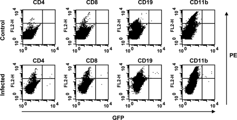
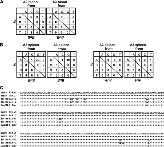
A novel gammaretrovirus, xenotropic murine leukemia virus-related virus (XMRV), has been identified in patients with prostate cancer and in patients with chronic fatigue syndromes. Standard Mus musculus laboratory mice lack a functional XPR1 receptor for XMRV and are therefore not a suitable model for the virus. In contrast, Gairdner's shrew-mice (Mus pahari) do express functional XPR1. To determine whether Mus pahari could serve as a model for XMRV, primary Mus pahari fibroblasts and mice were infected with cell-free XMRV. Infection of cells in vitro resulted in XMRV Gag expression and the production of XMRV virions. After intraperitoneal injection of XMRV into Mus pahari mice, XMRV proviral DNA could be detected in spleen, blood, and brain. Intravenous administration of a green fluorescent protein (GFP) vector pseudotyped with XMRV produced GFP(+) CD4(+) T cells and CD19(+) B cells. Mice mounted adaptive immune responses against XMRV, as evidenced by the production of neutralizing and Env- and Gag-specific antibodies. Prominent G-to-A hypermutations were also found in viral genomes isolated from the spleen, suggesting intracellular restriction of XMRV infection by APOBEC3 in vivo. These data demonstrate infection of Mus pahari by XMRV, potential cell tropism of the virus, and immunological and intracellular restriction of virus infection in vivo. These data support the use of Mus pahari as a model for XMRV pathogenesis and as a platform for vaccine and drug development against this potential human pathogen.


Figures

FIG. 1.
FIG. 1.
Mus pahari cells express functional receptor for XMRV. (A) Detection of XMRV proteins in primary Mus pahari cells by immunostaining. XMRV Gag and Env proteins (green) were detected by goat anti-MLV-p30/gp70 polyclonal antibodies. Cells and nuclei were counterstained with anti-β-actin antibody (red) and DAPI (blue), respectively. (B) Production of infectious XMRV virions from XMRV-infected Mus pahari cells. 293T cells were infected with the supernatants harvested from XMRV-infected and uninfected Mus pahari cells at 1 and 5 days after infection. XMRV-specific proviral DNA copy numbers in 293T cells were determined by real-time PCR as described in Materials and Methods.
FIG. 2.
FIG. 2.
Detection of XMRV-infected cells in spleen. Splenocytes of mice infected with a GFP-expressing XMRV were stained with PE-conjugated CD4, CD8, CD19, and CD11b, and analyzed by flow cytometry. The results of a 30,000 cell count are shown. Splenocytes of uninfected mouse were used as controls.
FIG. 3.
FIG. 3.
Induction of XMRV-specific antibodies in XMRV-injected mice. (A) Serially diluted plasma (1:20, 1:40, 1:80, 1:160, 1:320, and 1:640) isolated from XMRV-infected and uninfected control mice were incubated with GFP-expressing XMRV. Three days after infection, GFP-positive cell populations were analyzed by flow cytometry. Representative data with plasma dilutions at 1:20 and 1:640 were shown. (B) Detection of XMRV Env and Gag proteins by Western blotting with plasma samples of XMRV-infected mice. Cell lysates from PC3 cells (XMRV antigen negative, which is indicated as “−”), and cell lysates from XMRV-infected PC3 cells (XMRV antigen positive; “+”) were used as antigens. Plasma samples isolated at various time points (weeks postinfection, indicated under each blot) were used. Cell lysates were also analyzed by anti-MLV p30/gp70 antibody as a positive control.
FIG. 4.
FIG. 4.
Sequence analysis of partial XMRV gag and env genes recovered from infected Mus pahari. (A) The numbers of mutations in the gag gene from blood samples of mice A2 (total of 5,830 bp sequenced) and A3 (total of 1,740 bp sequenced) were summarized. (B) Numbers of mutations in the gag and env genes from spleen samples of mice A2 and A5 (in 5,830 bp for both gag sequenced and 1,785 bp for both env sequenced) were summarized. (C) Alignment of MLV-like sequences (#B Brain-1 and -2) identified in the brain of uninfected mouse #B was shown with corresponding env sequences of XMRV (22Rv1 and PCA17) and MLV CasE#1.

Similar articles

Cited by

References

    1. Battini, J. L., J. E. Rasko, and A. D. Miller. 1999. A human cell-surface receptor for xenotropic and polytropic murine leukemia viruses: possible role in G protein-coupled signal transduction. Proc. Natl. Acad. Sci. U. S. A. 96:1385-1390. - PMC - PubMed
    1. Caligiuri, M., et al. 1987. Phenotypic and functional deficiency of natural killer cells in patients with chronic fatigue syndrome. J. Immunol. 139:3306-3313. - PubMed
    1. Cloyd, M. W., M. M. Thompson, and J. W. Hartley. 1985. Host range of mink cell focus-inducing viruses. Virology 140:239-248. - PubMed
    1. D'Arcy, F., et al. 2008. No evidence of XMRV in Irish prostate cancer patients with the R462Q mutation. Eur. Urol. Suppl. 7:271.
    1. DesGroseillers, L., and P. Jolicoeur. 1984. Mapping the viral sequences conferring leukemogenicity and disease specificity in Moloney and amphotropic murine leukemia viruses. J. Virol. 52:448-456. - PMC - PubMed

Publication types

MeSH terms

LinkOut - more resources