. 2011 Jan;85(2):784-94.
doi: 10.1128/JVI.01397-10. Epub 2010 Nov 17.

The nuclear and adherent junction complex component protein ubinuclein negatively regulates the productive cycle of Epstein-Barr virus in epithelial cells

Affiliations

The nuclear and adherent junction complex component protein ubinuclein negatively regulates the productive cycle of Epstein-Barr virus in epithelial cells

Henri Gruffat et al. J Virol. 2011 Jan.

Abstract

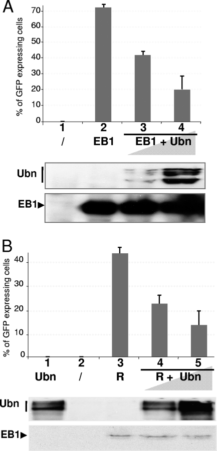
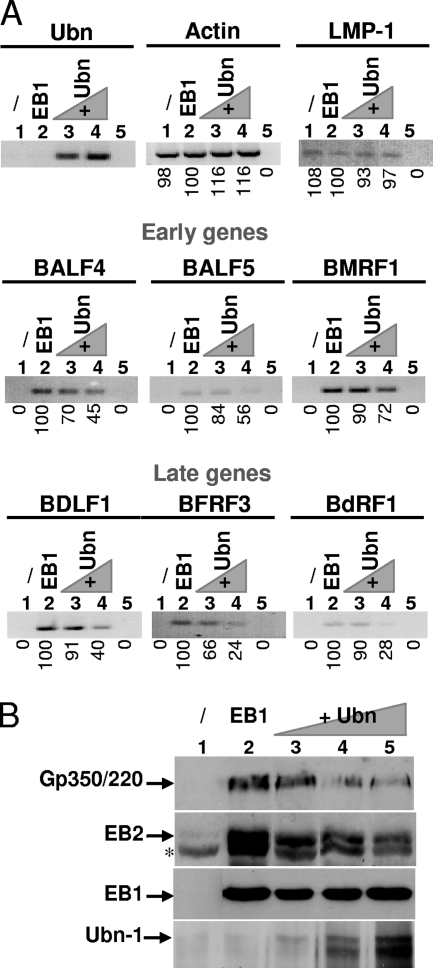
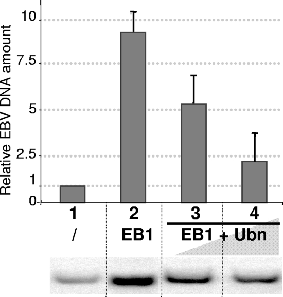
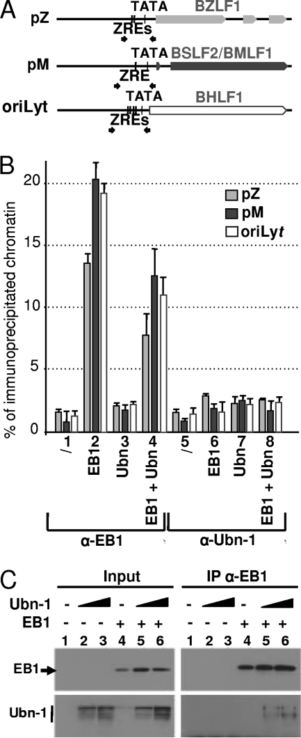
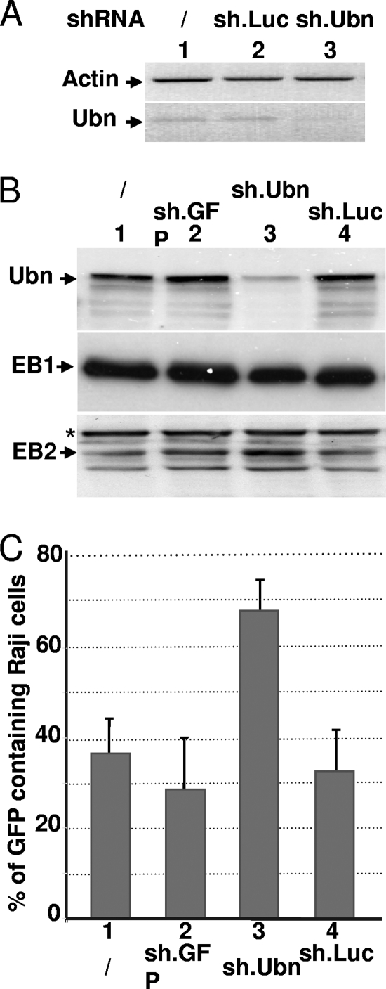
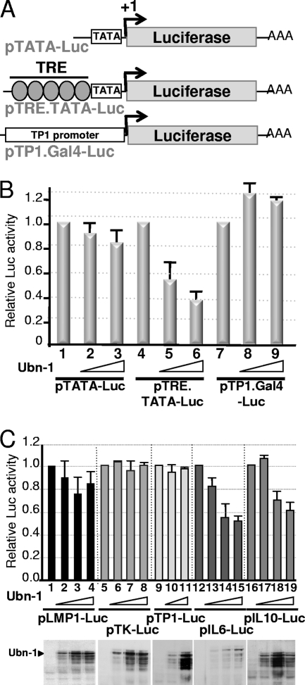
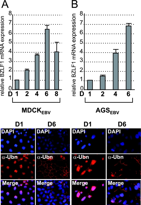
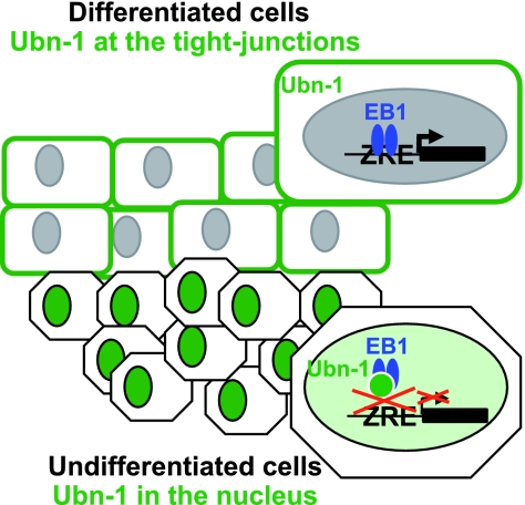
The Epstein-Barr Virus (EBV) productive cycle is initiated by the expression of the viral trans-activator EB1 (also called Zebra, Zta, or BZLF1), which belongs to the basic leucine zipper transcription factor family. We have previously identified the cellular NACos (nuclear and adherent junction complex components) protein ubinuclein (Ubn-1) as a partner for EB1, but the function of this complex has never been studied. Here, we have evaluated the consequences of this interaction on the EBV productive cycle and find that Ubn-1 overexpression represses the EBV productive cycle whereas Ubn-1 downregulation by short hairpin RNA (shRNA) increases virus production. By a chromatin immunoprecipitation (ChIP) assay, we show that Ubn-1 blocks EB1-DNA interaction. We also show that in epithelial cells, relocalization and sequestration of Ubn-1 to the tight junctions of nondividing cells allow increased activation of the productive cycle. We propose a model in which Ubn-1 is a modulator of the EBV productive cycle: in proliferating epithelial cells, Ubn-1 is nuclear and inhibits activation of the productive cycle, whereas in differentiated cells, Ubn-1 is sequestrated to tight junctions, thereby allowing EB1 to fully function in the nucleus.


Figures

FIG. 1.
FIG. 1.
Ubinuclein overexpression inhibits virion production in HEK293EBV cells. (A) Raji cells (2.5 × 10⁵) were incubated with 1 ml of filtered medium from HEK293EBV cells that had been either not transfected (lane 1), transfected with 0.5 μg of an EB1 expression plasmid (lane 2), or cotransfected with expression plasmids for both EB1 (0.5 μg) and increasing amounts of Ubn-1 (2 and 4 μg) (lanes 3 and 4). The amount of GFP-expressing Raji cells was determined by FACS analysis. The experiment was done 5 times, and the error bars represent standard deviations. (Lower panel) Immunoblot analysis for one representative experiment. Ubn-1 protein was detected using MAb ZAP1; EB1 protein was detected on the same membrane using MAb Z125. (B) Raji cells (2.5 × 10⁵) were incubated with 1 ml of filtered medium from HEK293EBV cells that had been either not transfected (lane 2), transfected with 4 μg of expression plasmid for Ubn-1 (lane 1) or 1 μg of an R expression plasmid (lane 3), or cotransfected with expression plasmids for both R (1 μg) and increasing amounts of Ubn-1 (2 and 4 μg) (lanes 3 and 4). The number of GFP-expressing Raji cells was determined by FACS analysis. (Lower panel) Immunoblot analysis for one representative experiment.
FIG. 2.
FIG. 2.
Ubinuclein modulates expression of both early and late EBV genes. (A) Total RNA from HEK293EBV cells that have been either not transfected (lane 1 of each panel), transfected with 0.5 μg of an EB1 expression plasmid (lane 2 of each panel), or cotransfected with expression plasmids for both EB1 (0.5 μg) and increasing amounts of Ubn-1 (2 and 4 μg) (lanes 3 and 4 of each panel) were purified, reverse transcribed, and analyzed by PCR using specific primers for actin (used to monitor the quality of mRNA purification and to normalize the amount of mRNA used under each condition), Ubn-1, the LMP-1 latent gene, the BALF4, BALF5, and BMRF1 early genes, and the BDLF1, BFRF3, and BdRF1 late genes. Lane 5 of each panel represents the results of a PCR amplification in the absence of reverse transcriptase. The results of PCR quantifications are indicated underneath each panel and are expressed relative to the amount of amplified DNA from the assay in which cells were transfected with an expression plasmid for EB1. (B) Immunoblot analysis of gp350/220, EB2, EB1, and Ubn-1 proteins expressed from HEK293EBV cells that have been either not transfected (lane 1), transfected with 0.5 μg of an EB1 expression plasmid (lane 2), or cotransfected with expression plasmids for both EB1 (0.5 μg) and increasing amounts of Ubn-1 (1, 2, and 4 μg) (lanes 3 and 4).
FIG. 3.
FIG. 3.
Ubinuclein overexpression inhibits viral DNA replication. Viral DNA extracted from HEK293EBV cells that had been either not transfected (lane 1), transfected with 0.5 μg of an EB1 expression plasmid (lane 2), or cotransfected with expression plasmids for both EB1 (0.5 μg) and increasing amounts of Ubn-1 (2 and 4 μg) (lanes 3 and 4) was quantified by Southern blotting using the BRRF1 probe. Fifty micrograms of total DNA obtained by Hirt extraction was cut by DpnI and BamHI and loaded in each lane. The results for one representative Southern blot are shown in the bottom panel. Quantification of the Southern blots is presented in the graph above as relative amounts of viral DNA observed after assignment of an arbitrary value of 1 to the quantified DNA present in the cells prior to reactivation of the productive cycle (lane 1). The experiment was done 3 times, and the error bars represent standard deviations.
FIG. 4.
FIG. 4.
Ubinuclein overexpression inhibits EB1-DNA interaction. (A) Schematic representation of the BZLF1 and BSLF2/BMLF1 promoters and those for the oriLyt region. TATA, TATA box; ZRE, EB1-responsive element. Small arrows indicate the positions of the primers used for the PCR. (B) HEK293EBV cells that had been either not transfected (lanes 1 and 5), transfected with 0.5 μg of an EB1 expression plasmid (lanes 2 and 6) or 4 μg of a Ubn-1 expression plasmid (lanes 3 and 7), or cotransfected with expression plasmids for both EB1 (0.5 μg) and Ubn-1 (4 μg) (lanes 4 and 8) were fixed with paraformaldehyde. The chromatin was then immunoprecipitated using either Z125 MAb directed against EB1 (lanes 1 to 4) or Zap-1 MAb directed against Ubn-1 (lanes 5 to 8). Immunoprecipitated DNA was then quantified by PCR using specific primers located on the pM or pZ promoter or on the oriLyt region. The results are given as the percentage of coimmunoprecipitated DNA relative to the amount of input DNA. The results obtained for the pZ promoter are shown in light gray, those for the pM promoter in dark gray, and those for the oriLyt region in white. (C) Coimmunoprecipitation of Ubn-1 and EB1 in HEK293EBV cells. HEK293EBV cells were transfected with 0.5 μg of EB1 expression plasmids or 2 and 4 μg of Ubn-1 expression plasmid as indicated in the figure. EB1 was immunoprecipitated using MAb Z125 and protein A magnetic beads (Millipore), and the immunoprecipitated proteins were analyzed by immunoblotting using either MAb Z125 or MAb ZAP1 to reveal the presence of EB1 or Ubn1, respectively (right panels). The total amount of each protein present in the extract prior to immunoprecipitation was evaluated by analysis of aliquots (1/10 for EB1 and 1/50 for Ubn-1) of cell extracts by immunoblotting as shown in the left panels.
FIG. 5.
FIG. 5.
Ubinuclein knockdown allows an increased production of virions. HEK293EBV cells were infected with lentiviruses coding for an Ubn-1-specific shRNA (sh.Ubn) or an unspecific shRNA, either sh.GFP or sh.Luc, and then transfected with an expression plasmid for EB1. The relative levels of both Ubn-1 mRNA and the actin control mRNA were measured by RT-PCR (A), and the relative amounts of Ubn-1, EB1, and BMLF1 proteins were evaluated by immunoblotting using specific antibodies (B). Supernatants from HEK293EBV cells infected with the shRNA-carrying lentiviruses and transfected by an EB1 expression vector were collected and used to infect Raji cells. The number of GFP-expressing Raji cells was determined by FACS analysis (C). The experiment was done 3 times, and the error bars represent standard deviations.
FIG. 6.
FIG. 6.
Ubinuclein interferes with AP1 trans-activation. (A) Schematic representation of the luciferase reporter constructs. TATA, TATA box; AAA, poly(A) site; TRE, AP-1-responsive element; +1, transcription initiation site. (B) HeLa cells were transfected with the various reporter constructs represented in panel A, together with no or increasing amounts of Ubn-1 expression plasmid. The luciferase activities presented in the histogram are given as relative values, using the results for the assay performed in the absence of Ubn-1 as a reference. (C) HeLa cells were transfected with reporter constructs carrying the luciferase-coding sequence under the control of various viral and cellular promoters as indicated in the figure, together with no or increasing amounts of Ubn-1 expression plasmid. The luciferase activities presented in the histogram are given as relative values, using the results for the assay performed in the absence of Ubn-1 as a reference. (Lower panel) Representative immunoblots from transfected HeLa cell extracts incubated with MAb ZAP1. The experiments were done 4 times, and the error bars represent standard deviations.
FIG. 7.
FIG. 7.
Correlation between ubinuclein localization and activation of the EBV productive cycle. Nonconfluent MDCKEBV cells (A) or AGSEBV cells (B) were plated in petri dishes. Cells were collected at different time points (D, day), and expression of the BZLF1 gene was monitored by semiquantitative RT-PCR. The results are presented as the amount of [32P]dCTP incorporation, relative to that obtained at day 1, which has been given an arbitrary value of 1. The experiments were done 4 times, and the error bars represent standard deviations. (Bottom panels) Localization of Ubn-1 was monitored by immunofluorescence staining at day 1 (nonconfluent cells) and day 6 (confluent cells). Cells were labeled using the anti-Ubn (α-Ubn) MAb ZAP1 (red); nuclei were visualized using DAPI (blue).
FIG. 8.
FIG. 8.
Model of control of the EBV productive cycle in epithelial cells. In undifferentiated cells, ubinuclein localized in the cell nucleus controls reactivation of the EBV productive cycle by inhibiting both AP1- and EB1-mediated gene expression, whereas in differentiated cells, ubinuclein is sequestered to the tight junctions so that EB1 is free to activate the productive cycle. ZRE, EB1-responsive elements.

Similar articles

Cited by

References

    1. Adamson, A. L., D. Darr, E. Holley-Guthrie, R. A. Johnson, A. Mauser, J. Swenson, and S. Kenney. 2000. Epstein-Barr virus immediate-early proteins BZLF1 and BRLF1 activate the ATF2 transcription factor by increasing the levels of phosphorylated p38 and c-Jun N-terminal kinases. J. Virol. 74:1224-1233. - PMC - PubMed
    1. Aho, S., M. Buisson, T. Pajunen, Y. W. Ryoo, J. F. Giot, H. Gruffat, A. Sergeant, and J. Uitto. 2000. Ubinuclein, a novel nuclear protein interacting with cellular and viral transcription factors. J. Cell Biol. 148:1165-1176. - PMC - PubMed
    1. Aho, S., J. Lupo, P. A. Coly, A. Sabine, M. Castellazzi, P. Morand, A. Sergeant, E. Manet, V. Boyer, and H. Gruffat. 2009. Characterization of the ubinuclein protein as a new member of the nuclear and adhesion complex components (NACos). Biol. Cell 101:319-334. - PubMed
    1. Babcock, G. J., L. L. Decker, M. Volk, and D. A. Thorley-Lawson. 1998. EBV persistence in memory B cells in vivo. Immunity 9:395-404. - PubMed
    1. Balaji, S., L. M. Iyer, and L. Aravind. 2009. HPC2 and ubinuclein define a novel family of histone chaperones conserved throughout eukaryotes. Mol. Biosyst. 5:269-275. - PMC - PubMed

Publication types

LinkOut - more resources