. 2011 Feb;85(3):1182-92.
doi: 10.1128/JVI.02143-10. Epub 2010 Nov 17.

Functional analysis of a novel motif conserved across geminivirus Rep proteins

Affiliations

Functional analysis of a novel motif conserved across geminivirus Rep proteins

Tara E Nash et al. J Virol. 2011 Feb.

Abstract

Members of the Geminiviridae have single-stranded DNA genomes that replicate in nuclei of infected plant cells. All geminiviruses encode a conserved protein (Rep) that catalyzes initiation of rolling-circle replication. Earlier studies showed that three conserved motifs-motifs I, II, and III-in the N termini of geminivirus Rep proteins are essential for function. In this study, we identified a fourth sequence, designated GRS (geminivirus Rep sequence), in the Rep N terminus that displays high amino acid sequence conservation across all geminivirus genera. Using the Rep protein of Tomato golden mosaic virus (TGMV AL1), we show that GRS mutants are not infectious in plants and do not support viral genome replication in tobacco protoplasts. GRS mutants are competent for protein-protein interactions and for both double- and single-stranded DNA binding, indicating that the mutations did not impair its global conformation. In contrast, GRS mutants are unable to specifically cleave single-stranded DNA, which is required to initiate rolling-circle replication. Interestingly, the Rep proteins of phytoplasmal and algal plasmids also contain GRS-related sequences. Modeling of the TGMV AL1 N terminus suggested that GRS mutations alter the relative positioning of motif II, which coordinates metal ions, and motif III, which contains the tyrosine involved in DNA cleavage. Together, these results established that the GRS is a conserved, essential motif characteristic of an ancient lineage of rolling-circle initiators and support the idea that geminiviruses may have evolved from plasmids associated with phytoplasma or algae.


Figures

FIG. 1.
FIG. 1.
Novel amino acid motif in the N termini of geminivirus replication proteins. (A) Amino acid sequence alignment shows the novel motif (designated GRS; dotted box) in the N termini of TGMV AL1 and Rep proteins of the type members of the begomovirus (Bean golden mosaic virus [BGMV]), curtovirus (Beet curly top virus [BCTV]), topocuvirus (Tomato pseudo-curly top virus [TPCTV]), and mastrevirus (Maize streak virus [MSV]) genera. Motifs I, II, and III, which are conserved among RCR initiator proteins (40), are marked by lines. The levels of identity for motif I (100%), motif II (67%), motif III (40%), and GRS (52%) are shown. Protein alignment was performed by using Vector NTI AlignX, and the color scheme represents amino acid identity (yellow), conservation (blue), and blocks of similarity (green) (45). (B) Amino acid sequence logo (13, 61) shows the conservation of the GRS motif across 198 geminivirus species (based on reference 21). The percent identity indicated below the sequence logo is based on the consensus amino acid at that position. Dots indicate residues in which the consensus sequence is shared among all genera. Amino acids are color coded according to their type as basic (blue), hydrophobic (black), polar/nonpolar (green), amide (purple), and acidic (red) (13, 61).
FIG. 2.
FIG. 2.
The GRS is required for TGMV infection and replication. (A) The positions of the alanine substitutions between TGMV AL1 amino acids 75 to 97 are indicated for mutants R-FD78, FH-N91, and IQ--K96. (B) N. benthamiana plants inoculated with a wild-type or mutant TGMV A replicon and a wild-type TGMV B replicon are shown. Total DNA from 5 plants of mock-, wild-type-, R-FD78-, FH-N91-, and IQ--K96-inoculated plants were analyzed for the presence of TGMV DNA-A by PCR (lanes 1 to 5 for each replicon). Lane C shows a control amplification with a wild-type TGMV A plasmid template. Lane M shows DNA size markers. (C) For transient replication assays, tobacco protoplasts were cotransfected with a wild-type TGMV B replicon and with plant expression cassettes encoding TGMV AL3 and wild-type AL1 (lane 1) or the mutants, R-FD78 (lane 2), FH-N91 (lane 3), or IQ--K96 (lane 4). Total DNA was isolated 48 h posttransfection and analyzed on DNA gel blots using a radiolabeled TGMV B probe. (D) For replication interference assays, tobacco protoplasts were cotransfected with a wild-type TGMV A replicon and with plant expression cassettes for R-FD78 (lane 3), FH-N91 (lane 4), or IQ--K96 (lane 5). The wild-type sample (lane 1) included an empty expression cassette. The AL1 mutant FQ118 (lane 2) was used as a positive control for interference (53). The graph shows the accumulation of TGMV A-DNA in the presence of the AL1 mutants relative to the empty cassette (wt, 100%). The error bars correspond to two standard errors. The asterisks indicate samples that are statistically different from wild-type (P < 0.05).
FIG. 3.
FIG. 3.
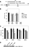
AL1 GRS mutants oligomerize and interact with other viral and host proteins. (A) Schematic of AL1 protein interactions (9, 38, 39, 62). (B) For AL1 oligomerization assays, yeast was cotransformed with a wild-type or mutant AL1 expression cassette fused to the GAL4 DBD wild-type (wt) or a mutant AL1 expression cassette fused to the GAL4 AD. Protein interactions were assayed by measuring β-galactosidase activity in soluble protein extracts. The error bars correspond to two standard errors. The asterisks indicate samples that are statistically different from the wild type (P < 0.05). (C) For AL1-protein interaction assays, yeast was cotransformed with a wild-type or mutant AL1 expression cassette fused to the GAL4 DBD and with AL3, RBR, PCNA, or GRIK expression cassettes fused to the GAL4 AD. Protein interactions were assayed as described in panel B. (D) Total proteins were extracted from yeast transformed with GAL4 DBD fusions corresponding to wild-type (lane 1), R-FD78 (lane 2), FH-N91 (lane 3), and IQ--K96 (lane 4) AL1. The extracts (100 μg) were fractionated by SDS-PAGE and analyzed by immunoblotting with an anti-GAL4 DBD antibody.
FIG. 4.
FIG. 4.
The GRS is required for DNA cleavage but not for DNA binding. (A) His-tagged TGMV AL11-180 proteins were expressed in E. coli and partially purified by Ni2+ affinity chromatography. Eluted proteins (2.5 μg) for wild-type AL1 (lane 1), R-FD78 (lane 2), FH-N91 (lane 3), and IQ--K96 (lane 4) were resolved by SDS-PAGE and visualized by Coomassie brilliant blue staining. (B) The dsDNA binding activities of wild-type AL11-180 (lanes 2 to 5) and the mutants R-FD78 (lanes 6 to 9), FH-N91(lanes 10 to 13), and IQ--K96 (lanes 14 to 17) were analyzed in EMSAs using a fluorescent dsDNA oligonucleotide (23). Lane 1 is a no protein control. All reactions contained a 500-fold excess of an unlabeled mutant dsDNA oligonucleotide and increasing amounts (0, 10×, 100×, or 500×) of unlabeled wild-type dsDNA oligo. Specific AL1-dsDNA complexes are designated “a” and “b”. (C) The ssDNA binding activities of wild-type AL11-180 (lanes 1 to 4) and the mutants R-FD78(lanes 5 to 8), FH-N91(lanes 9 to 12), and IQ--K96 (lanes 13 to 16) were analyzed in EMSAs using a fluorescent ssDNA oligonucleotide and increasing amounts (0, 10×, 50×, or 100×) of an unlabeled ssDNA oligonucleotide competitor. An AL1-ssDNA complex is marked by the dot. (D) The DNA cleavage activities of wild-type AL11-180 (lanes 2 and 7) and the mutants R-FD78 (lanes 3 and 8), FH-N91(lanes 4 and 9), and IQ--K96 (lanes 5 and 10) were analyzed using a fluorescent ssDNA oligonucleotide. Lanes 1 to 5 contained a wild-type ssDNA oligonucleotide, whereas lanes 6 to 10 contained a mutant oligonucleotide modified at the cleavage site, which cannot be cleaved. Lanes 1 and 6 correspond to no protein controls, whereas lane M contains a fluorescent oligonucleotide marker for the cleavage product. The dot indicates the cleaved oligonucleotide product. The faint double bands observed in lanes 4 and 9 are due to contaminating nonspecific nuclease activity. The DNA oligonucleotides used in panels B, C, and D are shown in Fig. S2B in the supplemental material.
FIG. 5.
FIG. 5.
(A) Alignment of TGMV AL1 amino acids 58 to 108, along with Rep proteins from Eragrostis curvula streak virus (ECSV), MSV, three phytoplasmids, and one algal plasmid (the accession numbers are shown). The GRS is designated by the dotted box, and dots above the alignment indicate positions of the GRS consensus residues. The protein alignment was performed by using Vector NTI AlignX, and the color scheme represents amino acid identity (yellow), conservation (blue), and blocks of similarity (green) (45). (B) Alignment of the structurally characterized Rep proteins from Tomato yellow leaf curl Sardinia virus (TYLCSV [8]), Faba bean necrotic yellows virus (FBNYV [74]), and Porcine circovirus type 2 (PCV2 [73]). The histidines in motif II and the catalytic tyrosine in motif III are marked by dots. Known structural elements are shown above the alignment for TYLCSV and below the alignment for FBNYV and PCV2 (75). (C) Model structure of TGMV AL1 amino acids 7 to 121. A PyMOL-generated protein ribbon model of TGMV AL1 based on the solved the 4-121 amino acid structure of TYLCSV Rep (PID 1L2M) (8) is shown. Selected residues in motif I (F16 and L17), motif II (H58 and H60), and motif III (Y104) are labeled. The inset box shows a rotated view including residues 8 Å from L17. The red mesh sphere shows the center of a hydrophobic cavity that could be created by mutating F77. The numbers indicate the angstrom distances from L17 to F77 or I92. The arrow shows a potential loop sequence between β6 and β7 strands (the amino acid numbering is for TGMV AL1, which is shifted by one position relative to the TYLCSV Rep structure [8] that served as the basis of the model). (D) The TGMV AL17-121 model was superimposed onto protein chains A and B of the SV40 T antigen bound to its cognate palindromic dsDNA (pdb code 2ITL). This model shows the positive protein surface of TGMV AL17-121 interacting with dsDNA. The electrostatic surface of the protein was created by using the Adaptive Poisson-Boltzmann Solver in PyMOL. In this model, R75 and K96 could contribute to dsDNA binding.

Similar articles

Cited by

References

    1. Ach, R. A., et al. 1997. RRB1 and RRB2 encode maize retinoblastoma-related proteins that interact with a plant D-type cyclin and geminivirus replication protein. Mol. Cell. Biol. 17:5077-5086. - PMC - PubMed
    1. Arguello-Astorga, G. R., and R. Ruiz-Medrano. 2001. An iteron-related domain is associated to motif 1 in the replication proteins of geminiviruses: identification of potential interacting amino acid base pairs by a comparative approach. Arch. Virol. 146:1465-1485. - PubMed
    1. Arnold, K., L. Bordoli, J. Kopp, and T. Schwede. 2006. The SWISS-MODEL workspace: a web-based environment for protein structure homology modeling. Bioinformatics 22:195-201. - PubMed
    1. Baase, W. A., L. Liu, D. E. Tronrud, and B. W. Matthews. 2010. Lessons from the lysozyme of phage T4. Protein Sci. 19:631-641. - PMC - PubMed
    1. Bagewadi, B., S. Chen, S. K. Lal, N. R. Choudhury, and S. K. Mukherjee. 2004. PCNA interacts with Indian mung bean yellow mosaic virus Rep and downregulates Rep activity. J. Virol. 78:11890-11903. - PMC - PubMed

Publication types

MeSH terms

LinkOut - more resources