Review
. 2010 Nov 17;30(46):15337-57.
doi: 10.1523/JNEUROSCI.3467-10.2010.

Metzincin proteases and their inhibitors: foes or friends in nervous system physiology?

Affiliations
Review

Metzincin proteases and their inhibitors: foes or friends in nervous system physiology?

Santiago Rivera et al. J Neurosci. .

Abstract

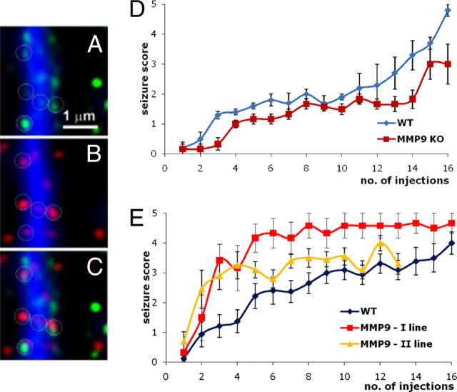
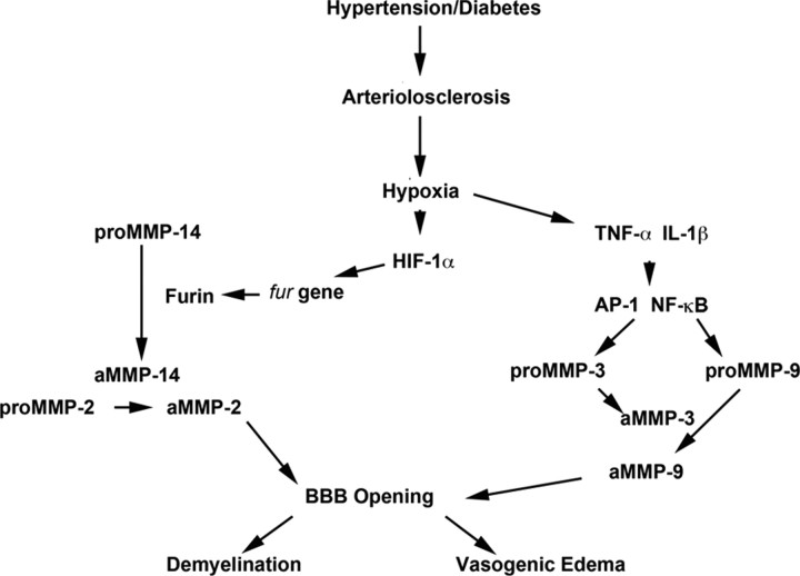
Members of the metzincin family of metalloproteinases have long been considered merely degradative enzymes for extracellular matrix molecules. Recently, however, there has been growing appreciation for these proteinases and their endogenous inhibitors, tissue inhibitors of metalloproteinases (TIMPs), as fine modulators of nervous system physiology and pathology. Present all along the phylogenetic tree, in all neural cell types, from the nucleus to the synapse and in the extracellular space, metalloproteinases exhibit a complex spatiotemporal profile of expression in the nervous parenchyma and at the neurovascular interface. The irreversibility of their proteolytic activity on numerous biofactors (e.g., growth factors, cytokines, receptors, DNA repair enzymes, matrix proteins) is ideally suited to sustain structural changes that are involved in physiological or postlesion remodeling of neural networks, learning consolidation or impairment, neurodegenerative and neuroinflammatory processes, or progression of malignant gliomas. The present review provides a state of the art overview of the involvement of the metzincin/TIMP system in these processes and the prospects of new therapeutic strategies based on the control of metalloproteinase activity.


Figures

Figure 1.
Figure 1.
Protease classification and structure. A, The human degradome, the repertoire of proteases produced by cells, consists of at least 569 proteases and homologs subdivided into five classes: 21 aspartic, 28 threonine, 150 cysteine, and 176 serine proteases and 194 metalloproteases, including MMP, ADAM, and ADAMTS family members. B, Structural classification of MMPs based on domain composition, including secreted and membrane-associated MMPs, and MMPs that are activated intracellularly via furin-mediated cleavage. C, Most ADAMs are type I transmembrane protein that possess disintegrin, cysteine-rich, and EGF domains in lieu of the MMP hemopexin domain. ADAMTSs are secreted proteins that contain thrombospondin I motifs in lieu of the EGF domain.
Figure 2.
Figure 2.
Physiopathological consequences of metzincin–substrate interactions. Nonexhaustive representation of interactions between metzincins and putative substrates in the nervous system, leading eventually to detrimental or beneficial effects in different physiological and pathological settings. The hierarchy between proteinase subtypes is established on the basis of current knowledge on metzincin actions and substrate preferences in the nervous system. The substrates include cytokines, soluble or ECM-bound growth factors (GF), and nuclear or membrane proteins. Metzincin-mediated proteolysis may lead to the following: (1) conversion of latent forms of proinflammatory cytokines (e.g., TNFα, IL-1β, etc.) or growth factors (e.g., BDNF, NGF) into their biologically active forms; (2) cleavage of nuclear (e.g., DNA repair enzymes) or ECM proteins (e.g., CSPGs, laminin, tenascin) causing irreversible changes in their structure and function; (3) cleavage of membrane proteins leading to their activation or inactivation or to the release of soluble ectodomains with, in most cases, yet unknown biological activity.
Figure 3.
Figure 3.
MMP activity is expressed at synapses, and MMP-9 plays a role in aberrant plasticity subserving epileptogenesis. A, MMP (gelatinolytic) activity revealed by in situ zymography (green) along a dendrite (blue, MAP-2 antibody staining). B, Dendritic spines (red, antibody staining against drebrin) along a dendrite. C, Colocalization of gelatinolytic activity and spine marker. D, MMP-9 KO mice and their wild-type siblings were treated to chemical kindling (an epileptogenic process) by repeated intraperitoneal injections of pentylenetetrazole (35 mg/kg, every 2–3 d). Seizure score reflects increasing severity of convulsions from 1 to 5. Note that MMP-9 KO are less susceptible to epileptogenesis. E, Pentylenetetrazole kindling in wild-type (WT) and transgenic (TG) rats overexpressing an autoactivating form of MMP-9 in neurons; two lines of transgenic rats were generated and subjected to kindling. Note that the transgenic rats are more prone to the epileptogenesis (for details on A–C, see Gawlak et al., 2009; for details on E and D, see Wilczynski et al., 2008).
Figure 4.
Figure 4.
Role of MMP-mediated proteolysis in loss of BBB integrity. Schematic diagram of a potential mechanism for MMP-mediated demyelination in vascular cognitive impairment. Hypertension and/or diabetes causes arteriolosclerosis of the blood vessels. Hypoxic hypoperfusion of the deep white matter results in induction of HIF-1α and cytokines (TNF-α and IL-1β). HIF-1α induces the fur gene and the protein furin, which activates proMMP-14 to the active form (aMMP-14). Then, aMMP-14 activates proMMP-2 to the active form of MMP-2. Cytokines induce the AP-1 and nuclear factor κB (NF-κB) transcription factors to produce proMMP-3 and proMMP-9. Active MMP-3 activates proMMP-9. Thus, MMPs open the BBB, leading to vasogenic edema in the white matter and demyelination.
Figure 5.
Figure 5.
Role of metzincins in tumor invasion. A, Primary malignant brain tumors grow as masses with irregular borders and contain regions of necrosis (black) and angiogenesis. The greatest glioma treatment challenge is tumor cell invasion. The most common route of invasion is along white matter tracts, including the corpus callosum into the contralateral hemisphere (1). Cells also migrate along the basement membrane of blood vessels (2) and spread subpially (3). Although some cells form satellites around neurons (4), others will terminate their migration at the white–gray interface (5). Metzincin proteases play multiple roles in tumor progression, with some possessing suppressive functions. B, Nonexhaustive representation of metzincins and TIMPs expressed in various cell types. This presentation comes with several caveats. First, all cell types have the “potential” to express a broad spectrum of metzincins. Second, not all proteases have been profiled with reliable tools in all cell types. Third, exclusion from the table does not preclude expression by that cell type under different physiological or pathological states. Nonetheless, the table stresses the concept that metzincins/TIMPs are not only produced by tumor cells but also by resident parenchymal and stromal cells. Thus, chemotherapies must take into account the normal physiological functions within these cell types.

References

    1. Adair JC, Charlie J, Dencoff JE, Kaye JA, Quinn JF, Camicioli RM, Stetler-Stevenson WG, Rosenberg GA. Measurement of gelatinase B (MMP-9) in the cerebrospinal fluid of patients with vascular dementia and Alzheimer disease. Stroke. 2004;35:e159–e162. - PubMed
    1. Agrawal SM, Lau L, Yong VW. MMPs in the central nervous system: where the good guys go bad. Semin Cell Dev Biol. 2008;19:42–51. - PubMed
    1. Ahmed Z, Dent RG, Leadbeater WE, Smith C, Berry M, Logan A. Matrix metalloproteases: degradation of the inhibitory environment of the transected optic nerve and the scar by regenerating axons. Mol Cell Neurosci. 2005;28:64–78. - PubMed
    1. Ahmed Z, Mazibrada G, Seabright RJ, Dent RG, Berry M, Logan A. TACE-induced cleavage of NgR and p75NTR in dorsal root ganglion cultures disinhibits outgrowth and promotes branching of neurites in the presence of inhibitory CNS myelin. FASEB J. 2006;20:1939–1941. - PubMed
    1. Akiguchi I, Tomimoto H, Suenaga T, Wakita H, Budka H. Blood-brain barrier dysfunction in Binswanger's disease; an immunohistochemical study. Acta Neuropathol (Berl) 1998;95:78–84. - PubMed

Publication types

MeSH terms