Comparative Study
. 2010 Nov 17;30(46):15358-68.
doi: 10.1523/JNEUROSCI.1862-10.2010.

Three Drosophila liprins interact to control synapse formation

Affiliations
Comparative Study

Three Drosophila liprins interact to control synapse formation

Sergio Astigarraga et al. J Neurosci. .

Abstract

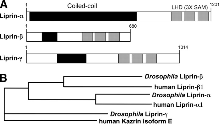
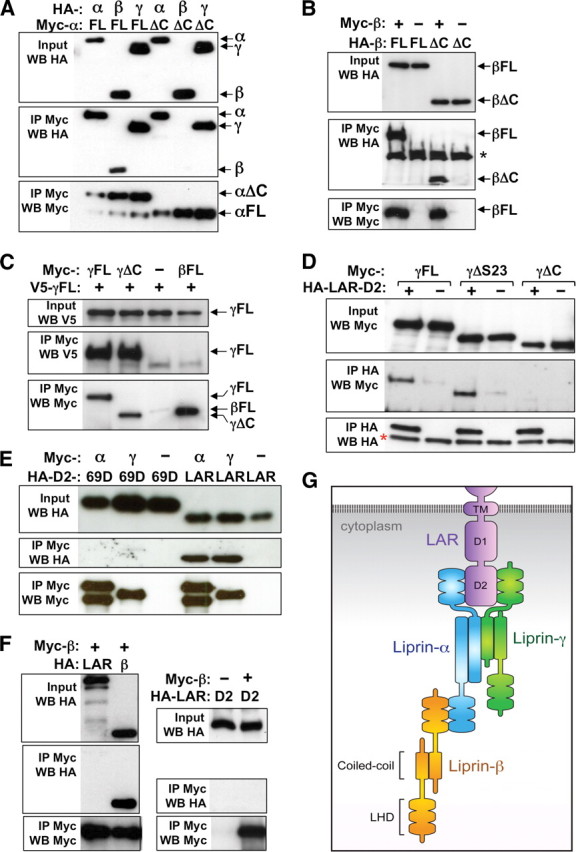
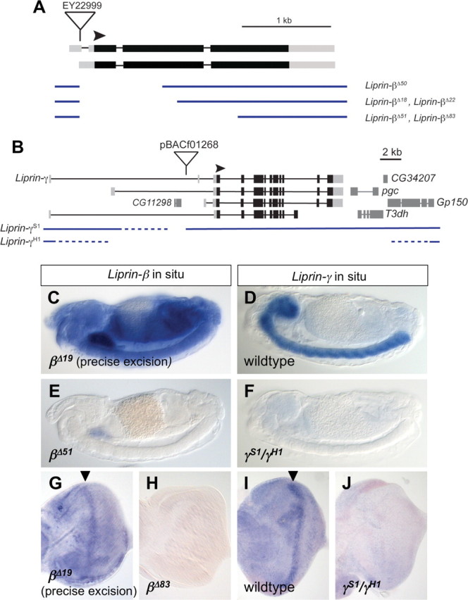
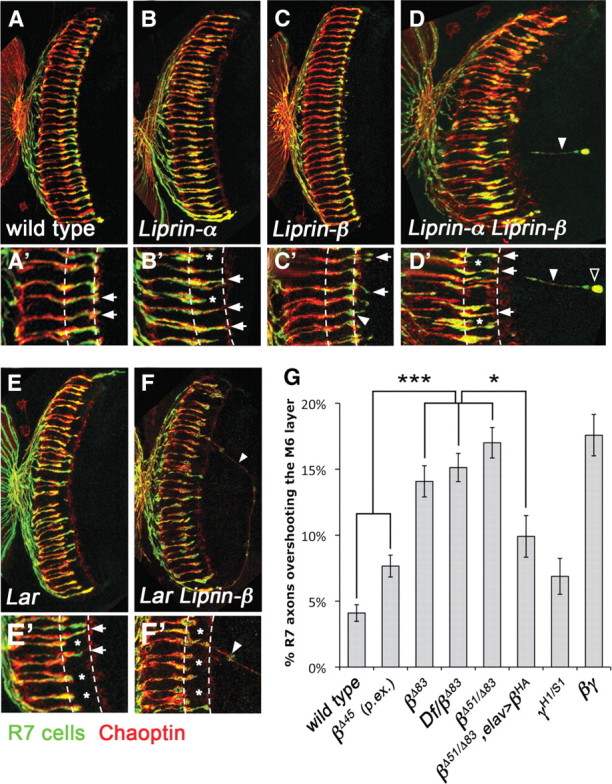
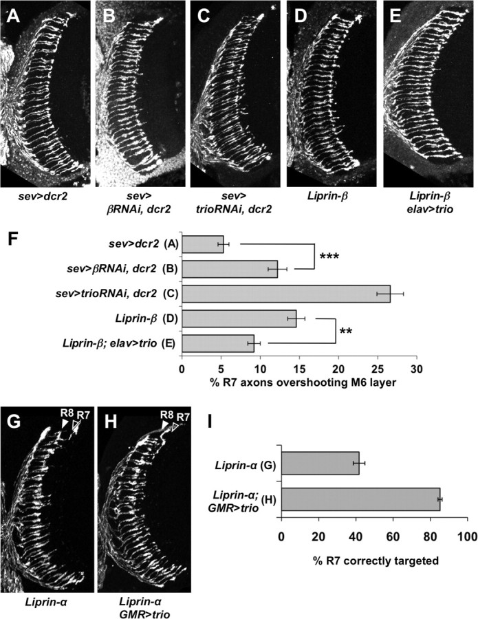
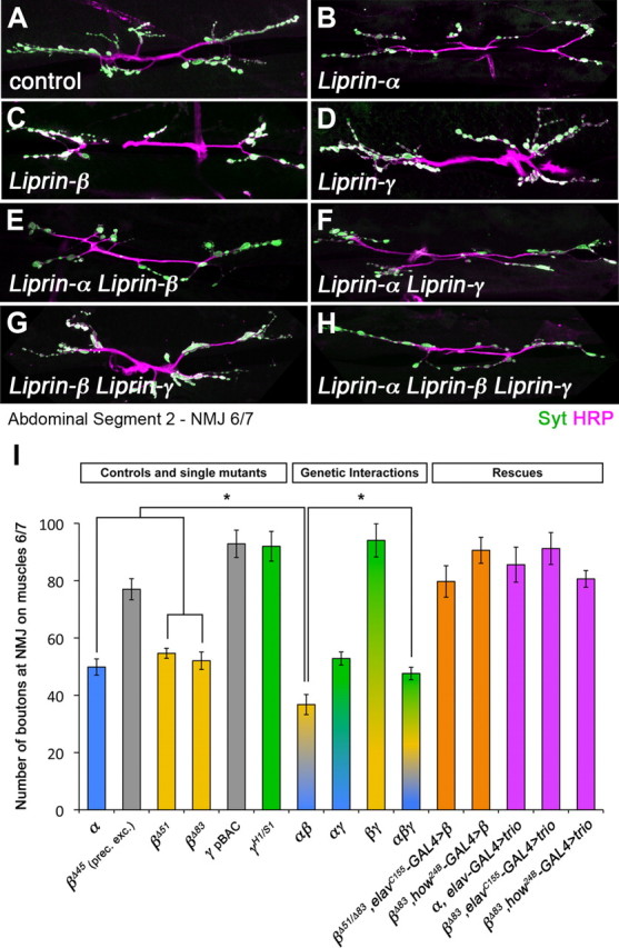
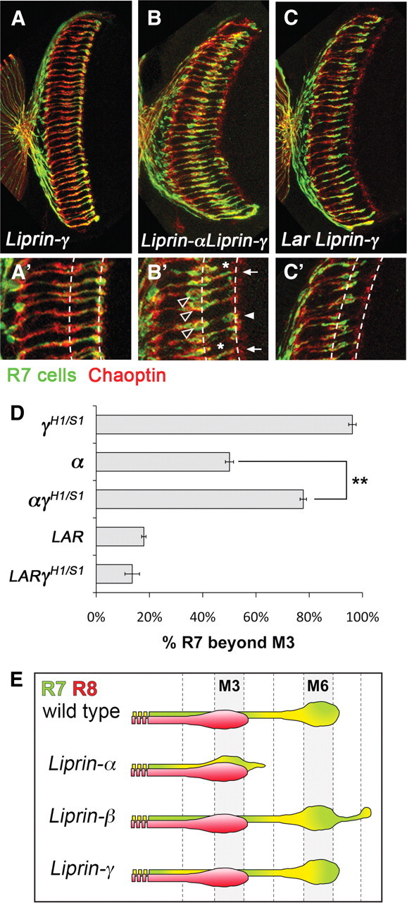
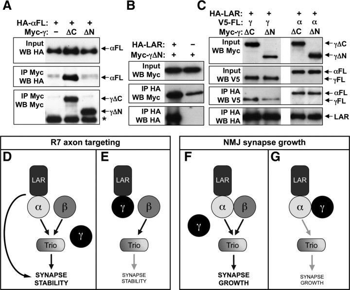
Liprin-α proteins are adaptors that interact with the receptor protein tyrosine phosphatase leukocyte common antigen-related (LAR) and other synaptic proteins to promote synaptic partner selection and active zone assembly. Liprin-β proteins bind to and share homology with Liprin-α proteins, but their functions at the synapse are unknown. The Drosophila genome encodes single Liprin-α and Liprin-β homologs, as well as a third related protein that we named Liprin-γ. We show that both Liprin-β and Liprin-γ physically interact with Liprin-α and that Liprin-γ also binds to LAR. Liprin-α mutations have been shown to disrupt synaptic target layer selection by R7 photoreceptors and to reduce the size of larval neuromuscular synapses. We have generated null mutations in Liprin-β and Liprin-γ to investigate their role in these processes. We find that, although Liprin-α mutant R7 axons terminate before reaching the correct target layer, Liprin-β mutant R7 axons grow beyond their target layer. Larval neuromuscular junction size is reduced in both Liprin-α and Liprin-β mutants, and further reduced in double mutants, suggesting independent functions for these Liprins. Genetic interactions demonstrate that both Liprin proteins act through the exchange factor Trio to promote stable target selection by R7 photoreceptor axons and growth of neuromuscular synapses. Photoreceptor and neuromuscular synapses develop normally in Liprin-γ mutants; however, removing Liprin-γ improves R7 targeting in Liprin-α mutants, and restores normal neuromuscular junction size to Liprin-β mutants, suggesting that Liprin-γ counteracts the functions of the other two Liprins. We propose that context-dependent interactions between the three Liprins modulate their functions in synapse formation.


Figures

Figure 1.
Figure 1.
The Drosophila Liprin family. A shows a schematic of the structures of the three Drosophila Liprin proteins. Predicted coiled-coil domains (amino acids 32–680 in Liprin-α, amino acids 93–185 in Liprin-β, and amino acids 173–352 in Liprin-γ) are depicted by black boxes, and the three C-terminal SAM domains that constitute the LHD (amino acids 942–1186 in Liprin-α, amino acids 359–602 in Liprin-β, and amino acids 582–816 in Liprin-γ) are shaded. B shows a phylogram generated by ClustalW of the evolutionary distances between the three Drosophila Liprins and their closest human homologs. Liprin-γ is equally homologous to both the other Drosophila Liprins, and most closely related to a specific isoform of the human Kazrin protein. Homologs of the other Kazrin isoforms, which do not contain the LHD, have not been reported in Drosophila.
Figure 2.
Figure 2.
Liprin-β and Liprin-γ homodimerize and bind to Liprin-α. A–E show coimmunoprecipitations of epitope-tagged proteins expressed in S2R+ cells. In A, HA-tagged Liprin-α, Liprin-β, or Liprin-γ was pulled down with Myc-tagged full-length (FL) Liprin-α or Liprin-α lacking the LHD (ΔC) by IP with anti-Myc. The middle panel shows the IP blotted (WB) with anti-HA, the top panel shows the input lanes blotted with anti-HA, and the bottom panel shows the IP blotted with anti-Myc. Both forms of Liprin-α pulled down Liprin-α and Liprin-γ, but only full-length Liprin-α pulled down Liprin-β. B, HA-tagged full-length (FL) Liprin-β, or Liprin-β lacking the LHD (ΔC), were pulled down with Myc-tagged Liprin-β by immunoprecipitation with anti-Myc. Liprin-β pulled down both forms of Liprin-β. The panels are labeled as in A. *Nonspecific band. C, V5-tagged Liprin-γ was pulled down with Myc-tagged full-length (FL) Liprin-γ, Liprin-γ lacking the LHD (ΔC), or full-length Liprin-β by immunoprecipitation with anti-Myc. Both forms of Liprin-γ, but not Liprin-β, could pull down Liprin-γ. D, Myc-tagged full-length (FL) Liprin-γ, or Liprin-γ lacking the last two SAM domains (ΔS23) or the whole LHD (ΔC) were pulled down with the HA-tagged D2 domain of LAR by immunoprecipitation with anti-HA. LAR-D2 pulled down Liprin-γ and Liprin-γΔS23, but not Liprin-γΔC, implicating the first SAM domain of Liprin-γ in the interaction with LAR. *Antibody heavy chain. E, The HA-tagged D2 domains of PTP69D (69D) or LAR were pulled down with Myc-tagged Liprin-α or Liprin-γ by immunoprecipitation with anti-Myc. Liprin-α and Liprin-γ pulled down LAR but not PTP69D. F, HA-tagged full-length LAR, LAR-D2, or Liprin-β were pulled down with Myc-tagged Liprin-β by immunoprecipitation with anti-Myc. Liprin-β pulled down Liprin-β but not LAR or the LAR D2 domain. In all panels, control lanes lacking any protein with the epitope tag recognized by the immunoprecipitating antibody are indicated by “−.” G shows a schematic of the interactions detected in this study and others. Liprin-α and Liprin-γ can bind to the D2 domain of LAR through their C-terminal SAM domains (Serra-Pagès et al., 1998). Liprin-α and Liprin-β interact through their LHDs (Serra-Pagès et al., 1998), and Liprin-α and Liprin-γ interact through the coiled-coil domain of Liprin-α. All three Liprins can homodimerize through their N-terminal coiled-coil domains (Serra-Pagès et al., 1998; Hofmeyer et al., 2006).
Figure 3.
Figure 3.
Generation of Liprin-β and Liprin-γ null mutants. A and B show the genomic structures of Liprin-β (A) and Liprin-γ (B). Exons are indicated by thick lines with untranslated regions in gray and translated regions in black, and introns by thin lines. The arrowheads indicate the start codons. Deletions induced by imprecise excision of P{EP}EY22999 (A) or by X-ray mutagenesis (B) are shown below (blue lines). The endpoints of the deletions in B have not been precisely mapped; the dotted lines indicate the regions within which they fall. C–J show in situ hybridization to stage 14 embryos (C–F) or third-instar eye imaginal discs (G–J) with Liprin-β (C, E, G, H) or Liprin-γ (D, F, I, J) probes. C, G, Precise excision of Liprin-β (LiprinΔ19). D, I, Wild type. E, LiprinΔ51. H, LiprinΔ83. F, J, LiprinS1/LiprinH1. The arrowheads in G and I indicate the morphogenetic furrow. Liprin-β is broadly expressed and Liprin-γ is specific to the nervous system in the embryo. Expression of both genes is abolished in the corresponding deletion mutants.
Figure 4.
Figure 4.
Liprin-β contributes to the termination of R7 axon growth in the correct target layer. A–F show head sections of adult flies. R7 and R8 axons are stained with anti-Chaoptin (red) and R7 axons are stained with anti-β-galactosidase to reveal the expression of panR7-lacZ (green). A′–F′ show enlargements of the medulla neuropil. The M3 and M6 layers are marked by dashed lines (M3, left; M6, right). A, A′, In wild-type flies, R7 photoreceptors project their axons to the M6 layer in the medulla and form round termini (arrows). B, B′, In Liprin-α mutants, R7 axons often terminate prematurely in M3, the R8 target layer (asterisks). Mutant R7 axons that reach the M6 layer usually do not expand their termini (arrows). C, C′, In Liprin-β mutants, some R7 axons extend beyond the M6 layer (arrows), even when the axons have normally expanded termini (arrowheads). D, D′, Many R7 axons in LiprinLiprin-β double mutants either do not reach their normal target layer (asterisks) or fail to expand their termini (arrows). Occasional R7 axons project much deeper into the brain (arrowhead), where they can form round termini (empty arrowhead). E, E′, Lar mutants show a stronger phenotype than Liprin-α; the majority of R7 axons stop in the M3 layer (asterisks). F, F′, In Lar Liprin-β double mutants, most R7 axons stop at the M3 layer (asterisks), but some project much further into the brain (F, F′, arrowhead). G, Quantification of the number of R7 axons that project beyond the M6 layer. Liprin-β mutants [LiprinΔ83: 14.08 ± 1.18%, 19 hemispheres (hm); Df(3L)BSC614/LiprinΔ83: 15.13 ± 1.07%, 15 hm; and LiprinΔ51/LiprinΔ83: 17.01 ± 1.16%, 36 hm] have more protrusions than the controls (wild type: 4.10 ± 0.63%, 14 hm; precise excision LiprinΔ45: 7.66 ± 0.83%, 24 hm). This phenotype is rescued by the expression of a UAS-Liprin-β transgene with the pan-neuronal driver elav-GAL4 (9.91 ± 1.58%, 13 hm). Liprin-γ mutants (6.88 ± 1.36%, 10 hm) are comparable with the control, and LiprinLiprin-γ double mutants (17.58 ± 1.57%, 7 hm) are similar to Liprin-β. Error bars represent SEM. *p < 0.05; ***p < 0.0001.
Figure 5.
Figure 5.
Trio acts downstream of Liprin-β and Liprin-α in R7 targeting. A–E show adult head sections stained with anti-β-galactosidase to reveal glass-lacZ expression. A, UAS-dcr2/CyO; sev-GAL4/TM6c (control). B, UAS-dcr2/+; sev-GAL4/UAS-LiprinRNAi. C, UAS-dcr2/+; sev-GAL4/UAS-trio RNAi. D, LiprinΔ51. E, elav-trio/CyO; LiprinΔ51. F shows a quantification of the percentage of axons projecting beyond the M6 layer in each genotype. Expression of trio RNAi causes overshooting of the R7 target layer (26.6 ± 1.7%, 17 hm) similar to Liprin-β mutants (14.6 ± 1.1%, 18 hm) or expression of LiprinRNAi in R7 (12.2 ± 1.2%, 18 hm). Overexpression of Trio in a Liprin-β mutant background rescues the phenotype (9.2 ± 0.8%, 19 hm). G and H show adult head sections stained with anti-β-galactosidase to reveal glass-lacZ expression. G, Liprinoos. H, Liprinoos; GMR-trio. I shows a quantification of the percentage of axons projecting beyond the M3 layer in each genotype. Overexpression of Trio in a Liprin-α mutant background rescues the early termination of R7 axons (85.2 ± 1.1% R7 axons correctly targeted, 18 hm, 907 cl, compared with 41.7 + 3.1%, 10 hm, 646 cl for Liprin-α alone). cl, Columns (pairs of R7–R8). The arrows indicate the R7 and R8 target layers. Error bars represent SEM. **p < 0.001; ***p < 0.0001.
Figure 6.
Figure 6.
Neuromuscular synapse size is reduced in Liprin-β mutants. A–H show synapses formed on muscles 6 and 7 in abdominal segment 2 (A2) of third-instar larvae. Nerves are stained with anti-HRP (magenta) and synaptic boutons with anti-Synaptotagmin (green). A, Control synapse from pBACf01268, the line in which Liprin-γ mutations were generated. B, Liprinoos. C, LiprinΔ51. D, LiprinH1/LiprinS1. E, Liprinoos; LiprinΔ51. F, Liprinoos, LiprinH1/Liprinoos, LiprinS1. G, LiprinH1/LiprinS1; LiprinΔ51. H, Liprinoos, LiprinH1/Liprinoos, LiprinS1; LiprinΔ51. I, Quantification of the number of boutons in abdominal segment 2 for the genotypes indicated. The precise excision line Δ45 was used as a control for Liprin-β, and pBACf01268, which does not affect Liprin-γ expression, was used as a control for Liprin-γ. Liprin-α and Liprin-β mutants have fewer boutons than controls, and the LiprinLiprin-β double-mutant synapse is even smaller. Liprin-γ single mutants have normally sized synapses and removal of Liprin-γ does not affect Liprin-α mutants. However, removal of Liprin-γ increases the number of boutons at Liprin-β or LiprinLiprin-β mutant synapses. Liprin-α is rescued by expressing UAS-trio in neurons using elavC155-GAL4. Liprin-β is rescued by expressing UAS-Liprin-β or UAS-trio either in neurons, using elavC155-GAL4, or in muscles, using how24B-GAL4. Between 14 and 23 NMJs were analyzed for each genotype. Error bars represent SEM. *p < 0.05.
Figure 7.
Figure 7.
Liprin-γ antagonizes Liprin-α in R7 targeting. A–C show adult head sections stained with anti-Chaoptin (red) and anti-β-galactosidase to reveal the expression of panR7-lacZ (green). Regions of the medulla neuropil are enlarged in A′–C′. A, A′, Liprin-γ mutants show normal R7 targeting. B, B′, Liprin-α Liprin-γ double mutants. More R7 axons reach the M6 layer than in Liprin-α single mutants. Interestingly, some R7 axons expand in the M3 layer (empty arrowheads), although they also form round termini in the M6 layer (arrowhead). C, C′, Lar Liprin-γ double mutants are similar to Lar single mutants. E shows a quantification of the percentage of R7 axons projecting beyond the M3 layer in the indicated genotypes. The Liprin-α phenotype (50.06% ± 1.52%, 21 hm, 1605 cl) is improved when Liprin-γ is removed in this background (77.77 ± 1.20%, 16 hm, 1588 cl), suggesting an antagonistic relationship between Liprin-α and Liprin-γ. However, the Lar phenotype (17.90 ± 0.82%, 15 hm, 1872 cl) is not rescued in Lar Liprin-γ double mutants (13.52 ± 2.74%, 9 hm, 533 cl). E shows a diagram of the R7 phenotypes of the three Liprin mutants. Error bars represent SEM. **p < 0.001.
Figure 8.
Figure 8.
Competitive interactions between Liprins and LAR. A–C show coimmunoprecipitations of epitope-tagged proteins expressed in S2R+ cells. A, Full-length HA-tagged Liprin-α is pulled down with Myc-tagged Liprin-γΔC or Liprin-γΔN by immunoprecipitation with anti-Myc. Input and IP lanes are blotted as indicated on the left. Liprin-γΔC, but not Liprin-γΔN, binds to Liprin-α. *Antibody heavy chain. B, Myc-tagged Liprin-γΔN is pulled down with HA-tagged LAR by immunoprecipitation with anti-HA. Liprin-γΔN binds to LAR. C, V5-tagged full-length Liprin-γ (left) or Liprin-α (right) is pulled down with HA-tagged LAR by immunoprecipitation with anti-HA in the presence of Myc-tagged Liprin-γΔC, a control protein that does not bind to LAR, or Myc-tagged Liprin-γΔN, which can bind to LAR. Input and IP lanes are blotted as indicated on the left. Liprin-γΔN reduces the amount of full-length Liprin-γ coimmunoprecipitated by LAR, indicating that the two forms of Liprin-γ compete for binding to LAR. However, the presence of Liprin-γΔN does not affect the interaction between Liprin-α and LAR. D shows a possible mechanism for R7 targeting, in which LAR, Liprin-α, and Liprin-β promote synapse stabilization at least in part through Trio. Loss of Liprin-β or Trio causes mutant R7 axons to project beyond the M6 layer rather than retracting to the M3 layer, suggesting that LAR and Liprin-α have additional downstream effectors. E, Liprin-γ may bind to and antagonize LAR in the absence of Liprin-α, contributing to the loss of synapse stability in Liprin-α mutants. F shows a possible mechanism for NMJ growth. Liprin-α and Liprin-β make independent contributions to synapse growth, again acting through Trio. G, Liprin-γ might bind to and antagonize Liprin-α in the absence of Liprin-β, contributing to the lack of synapse growth in Liprin-β mutants.

References

    1. Aberle H, Haghighi AP, Fetter RD, McCabe BD, Magalhães TR, Goodman CS. wishful thinking encodes a BMP type II receptor that regulates synaptic growth in Drosophila. Neuron. 2002;33:545–558. - PubMed
    1. Ball RW, Warren-Paquin M, Tsurudome K, Liao EH, Elazzouzi F, Cavanagh C, An BS, Wang TT, White JH, Haghighi AP. Retrograde BMP signaling controls synaptic growth at the NMJ by regulating Trio expression in motor neurons. Neuron. 2010;66:536–549. - PubMed
    1. Bateman J, Shu H, Van Vactor D. The guanine nucleotide exchange factor trio mediates axonal development in the Drosophila embryo. Neuron. 2000;26:93–106. - PubMed
    1. Biederer T, Stagi M. Signaling by synaptogenic molecules. Curr Opin Neurobiol. 2008;18:261–269. - PMC - PubMed
    1. Choe KM, Prakash S, Bright A, Clandinin TR. Liprin-α is required for photoreceptor target selection in Drosophila. Proc Natl Acad Sci U S A. 2006;103:11601–11606. - PMC - PubMed

Publication types

MeSH terms

Substances