Comparative Study
. 2010 Nov 17;30(46):15383-99.
doi: 10.1523/JNEUROSCI.2193-10.2010.

Processing afferent proprioceptive information at the main cuneate nucleus of anesthetized cats

Affiliations
Comparative Study

Processing afferent proprioceptive information at the main cuneate nucleus of anesthetized cats

Roberto Leiras et al. J Neurosci. .

Abstract

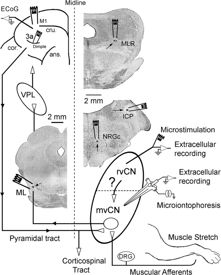
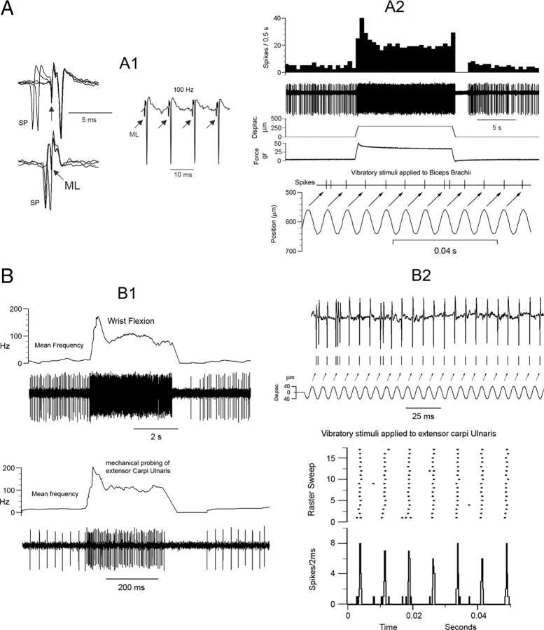
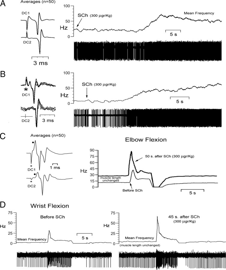
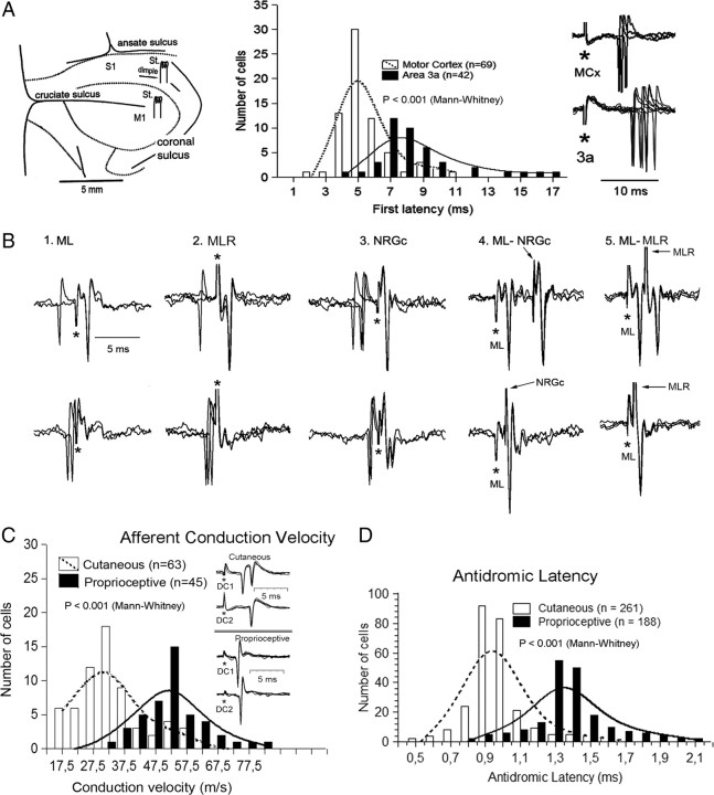
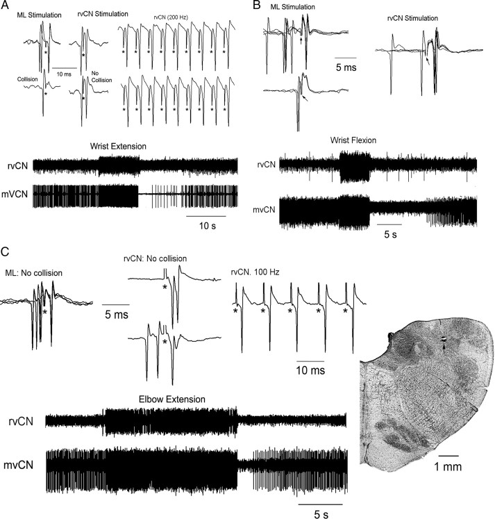
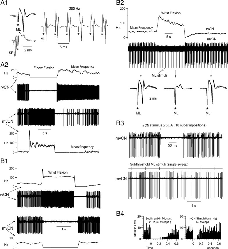
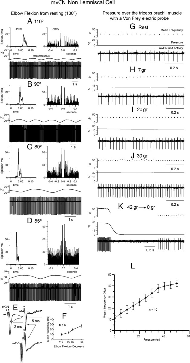
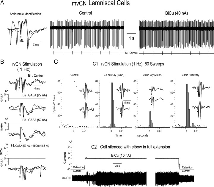
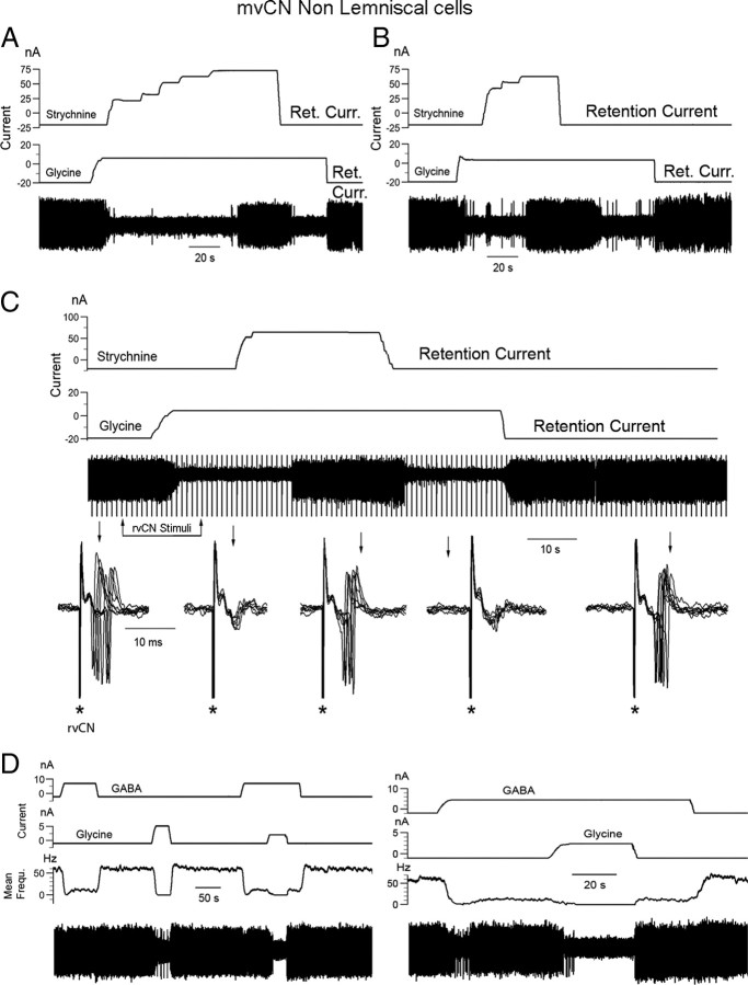
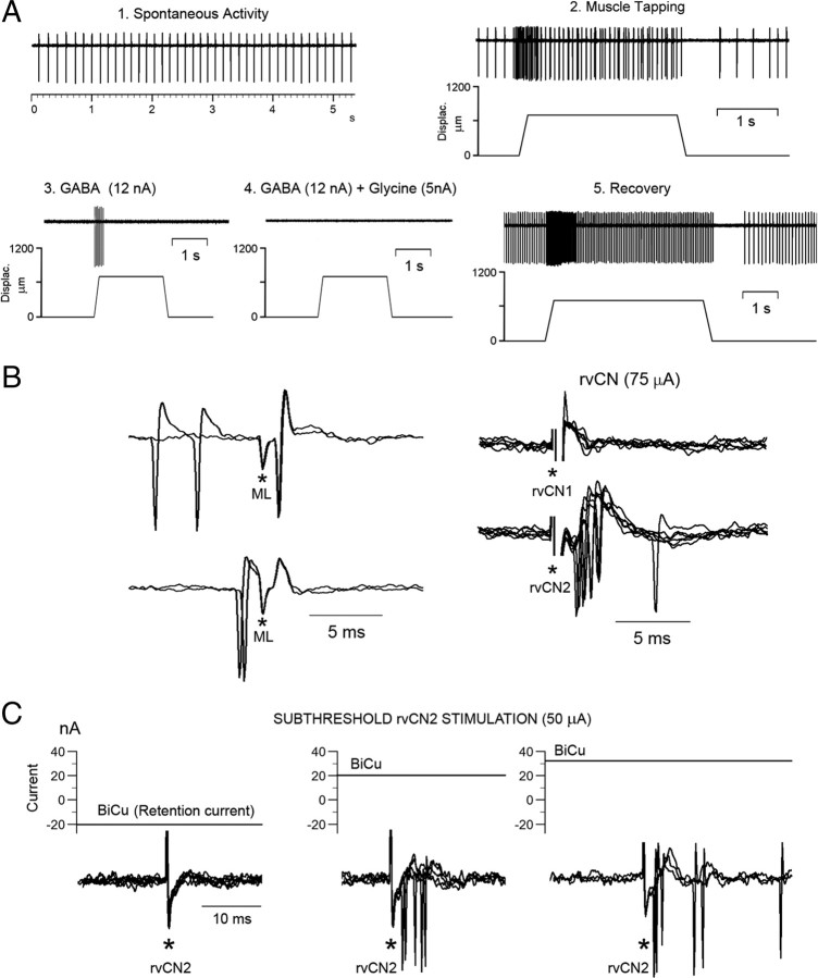
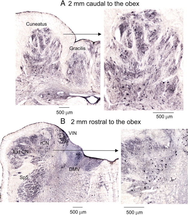
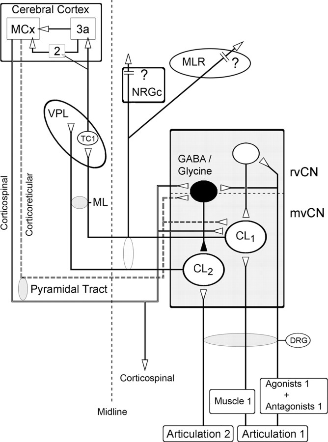
Medial lemniscal activity decreases before and during movement, suggesting prethalamic modulation, but the underlying mechanisms are largely unknown. Here we studied the mechanisms underlying proprioceptive transmission at the midventral cuneate nucleus (mvCN) of anesthetized cats using standard extracellular recordings combined with electrical stimulation and microiontophoresis. Dual simultaneous recordings from mvCN and rostroventral cuneate (rvCN) proprioceptive neurons demonstrated that microstimulation through the rvCN recording electrode induced dual effects on mvCN projection cells: potentiation when both neurons had excitatory receptive fields in muscles acting at the same joint, and inhibition when rvCN and mvCN cells had receptive fields located in different joints. GABA and/or glycine consistently abolished mvCN spontaneous and sensory-evoked activity, an effect reversed by bicuculline and strychnine, respectively; and immunohistochemistry data revealed that cells possessing strychnine-sensitive glycine receptors were uniformly distributed throughout the cuneate nucleus. It was also found that proprioceptive mvCN projection cells sent ipsilateral collaterals to the nucleus reticularis gigantocellularis and the mesencephalic locomotor region, and had slower antidromic conduction speeds than cutaneous fibers from the more dorsally located cluster region. The data suggest that (1) the rvCN-mvCM network is functionally related to joints rather than to single muscles producing an overall potentiation of proprioceptive feedback from a moving forelimb joint while inhibiting, through GABAergic and glycinergic interneurons, deep muscular feedback from other forelimb joints; and (2) mvCN projection cells collateralizing to or through the ipsilateral reticular formation allow for bilateral spreading of ascending proprioceptive feedback information.


Figures

Figure 1.
Figure 1.
General experimental arrangement. Stimulating electrodes were placed in the contralateral ML, the primary motor cortex (M1), and area 3a; and in the ipsilateral NRGc, inferior cerebellar peduncle (ICP), and MLR. Extracellular simultaneous recording was performed at the rvCN and mvCN. Microstimulation applied through the rvCN recording electrode served to test its effects on mvCN proprioceptive cells. Microiontophoretic ejection was used to test the effects exerted by GABA and glycine on the spontaneous and evoked activity of mvCN neurons. See text for further details. Three coronal sections show small electrolytic lesions (signaled by arrows) at the ML, MLR, NRGc, and ICP. ans, Ansate sulcus; cru, cruciate sulcus; cor, coronal sulcus; DRG, dorsal root ganglia; VPL, thalamic ventroposterolateral nucleus.
Figure 2.
Figure 2.
Characterization of proprioceptive mvCN neurons. Two different CL cells are illustrated (A, B). A1, Antidromic identification showing 1:1 high-frequency following to ML stimuli (right panel) and collision when ML stimuli were applied after spontaneous spikes (SP) at the adequate interval (left panel). The arrows point to the ML stimulus artifacts. A2, This neuron was activated by tapping the biceps brachii with a feedback mechanical stimulator at low and high frequencies. The records show the following (from top to bottom): discharge rate, unit activity, probe displacement, force applied to the muscle, discriminated spike responses, and sinusoidal probe vibration at 135 Hz. The arrows point to the spike responses produced by each sinusoid. B, A different CL cell was activated by wrist flexion and by tapping the extensor carpi ulnaris at low (B1) and high (B2) frequencies. Note in the histogram and corresponding raster plot of B2 the tight coupling between the positive sinusoidal peaks, beginning at time 0, and the evoked cellular activity. In the raster plot, each row represents a sweep during which 7 consecutive sinusoidal stimuli were applied, and each dot represents a spike.
Figure 3.
Figure 3.
Sensitivity of mvCN neurons to intravenous injection of SCh. A–D, Four different cells. A, B, Resting activity, with muscle length unchanged, was greatly increased following SCh injection. The insets at left show averages (A) and single-sweep superimpositions (B) of responses to two-point stimulation at the ipsilateral cervical dorsal column (DC1; DC2). C, The resting (static) and dynamic activity to elbow flexion of a different mvCN cell were increased after SCh injection. The inset at left shows the averages of 50 responses to two-point dorsal column stimulation with the black dots signaling positive field potentials reflecting the arrival of presynaptic volleys (Andersen et al., 1964a). D, Another mvCN cell showing a significant increase in activity during the dynamic phase of wrist flexion, while resting activity increased very little after SCh injection.
Figure 4.
Figure 4.
Motor cortex stimulation activated mvCN-CL cells at shorter latencies than area 3a. The mvCN projection neurons sent collateral branches to the ipsilateral NRGc and MLR, and showed shorter afferent and longer antidromic latencies than cutaneous CL neurons from the cluster region. A, Schematic drawing of stimulating electrodes placed in the cerebral cortex (left). The histograms illustrate the response latencies to motor cortex and area 3a stimulation with a sample of superimposed sweeps shown at right. The first latency data to motor cortex and area 3a stimulation well fit to two Gaussian distributions centered at 5 and 8 ms, respectively. B, Superimposed records illustrating a CL neuron sending a collateral branch to or through the NRGc and the MLR with the antidromic responses shown in the upper part and collision with spontaneous or evoked spikes in the lower part of each panel (1–3). Reciprocal ML-NRGc and ML-MLR collision tests are shown in panels 4 and 5, as indicated. Stimulus artifacts are marked by asterisks. In panel 5, the polarity of ML stimulation was reversed. C, Histogram showing the bimodal Gaussian distributions of cutaneous cells from the cluster region and mvCN proprioceptive neurons, grouped according to the conduction velocities of afferent fibers driving them (bins of 5 m/s). The single-sweep superimpositions at the inset show examples of afferent-induced responses generated on one cutaneous (top) and one proprioceptive (bottom) cells. D, Histograms of the antidromic latencies of cutaneous and proprioceptive CL cells recorded in the cluster region and mvCN, respectively. Their distribution is bimodal and well fit by two Gaussian graphs centered at 0.95 and 1.35 ms.
Figure 5.
Figure 5.
Dual recordings demonstrated that whenever mvCN and rvCN neurons had excitatory receptive fields at the same joint, rvCN microstimulation activated mvCN cells. The records illustrate the behavior of two CL cells (A, B) and one non-CL neuron (C). A, An ML-antidromic mvCN neuron (upper left, antidromic identification) responding to wrist extension was orthodromically activated by stimulation at an rvCN site also activated by wrist extension (lower records). Note that the mvCN cell faithfully followed 200 Hz rvCN repetitive stimulation, but collision did not occur (upper). B, Another CL neuron (upper left, antidromic identification) responding to wrist flexion was orthodromically activated (upper right, superimposed sweeps) by stimulating an rvCN site also activated by wrist flexion (lower records). C, A non-CL mvCN neuron (upper left, no collision) sensitive to elbow extension (lower) was orthodromically activated by stimulating at an rvCN site (upper, right two panels) also sensitive to elbow extension (lower). Note that the mvCN cell followed 100 Hz iterative rvCN stimulation, but collision did not occur (upper right). An electrolytic lesion (signaled by an arrow) produced at this stimulating rvCN site is shown at right. Stimulus artifacts are marked by asterisks.
Figure 6.
Figure 6.
When simultaneously recorded mvCN and rvCN neurons had receptive fields in distinct articulations, stimulation at the rvCN site inhibited the concurrently recorded mvCN cell. A, B, Two different cells. A, Antidromic identification of a CL neuron (A1) activated by elbow flexion (A2, lower). Elbow flexion inhibited another concurrently recorded rvCN cell (A2, upper). B, Wrist flexion activated an rvCN neuron and inhibited a simultaneously recorded mvCN-CL (B1). The antidromic spikes of the CL cell failed during wrist flexion (B2, superimposed sweeps expanded below), indicating that wrist flexion exerted a true inhibition on the mvCN neuron. Microstimulation at the rvCN recording site transitorily suppressed the activity of the mvCN cell (B3, upper). Subthreshold ML stimulation also transiently silenced the mvCN neuron (B3, lower). The peristimulus histograms in B4 (stimuli at time 0) show that the ML induced inhibition was longer lasting than the rvCN inhibition. Stimulus artifacts are marked with asterisks in A1 and the expanded records in B2; the stimulus artifacts in B3 were recorded on a different channel shown above each neuronal recording.
Figure 7.
Figure 7.
mvCN neurons were sensitive to muscle steady stretch. Same non-CL neuron. A–D, This non-CL cell showed a slow rhythmic activity at ∼0.4 Hz. The elbow joint was gradually flexed from rest (130°) and maintained at each new position for 5 min. The activity of the non-CL cell gradually incremented with lengthening of extensors. INTH, Interval interspike histogram; AUTO, autocorrelogram. E, This presumed interneuron responded antidromically to rvCN stimulation. The two superimpositions at left show the all or none nature of the antidromic spike and the superimposed records at right illustrate the collision test. F, Histogram (mean ± SEM) showing an almost linear increase in activity with elbow flexion for six different cells (4 CL). G–K, Increasing the steady pressure applied to the triceps brachii produced a similar increase in frequency as that observed with steady stretch. Note the firing silence at the end of stimulation (K). L, Histogram illustrating the mean ± SEM frequency increase with pressure for 10 different cells (7 CL).
Figure 8.
Figure 8.
GABA and glycine shape the responses of mvCN projection cells. A–C, Three different CL neurons. A, Antidromic identification (left) showing that collision occurred when a spontaneous spike preceded the antidromic response at the adequate interval. The single-sweep records show transient silenced firing following antidromic ML spikes (left sweep) that was abolished by BiCu ejection (right sweep). B, A different CL cell was orthodromically activated by rvCN stimulation (no collision in B1), and GABA ejection (52 nA) blocked the rvCN-induced excitation (B2, B3), an effect that was reversed by simultaneously ejecting BiCu (B4). C, Glycine blocked the rvCN-induced activation of another CL cell (C1). BiCu restored the cellular activity when suppressed by fully extending the elbow (C2). This cell increased firing when flexing the elbow.
Figure 9.
Figure 9.
Effects of glycine, strychnine, and GABA on mvCN presumed interneurons. A–D, Four different spontaneously active non-CL cells. A, B, The suppression of spontaneous activity by glycine was abolished by concurrent strychnine ejection. C, The synaptic activation produced by rvCN stimulation (see the superimposed records expanded below) was abolished by glycine ejection and restored when simultaneously ejecting strychnine. D, Additive effects on spontaneous activity by simultaneously ejecting GABA and glycine, as indicated.
Figure 10.
Figure 10.
GABA and glycine exerted additive inhibitory effects over proprioceptive induced responses, and BiCu ejection unmasked a GABAA-dependent tonic inhibition. A–C, Same CL cell. A, GABA application was adjusted to leave only part of the dynamic response when tapping the muscle with an electric Von Frey probe (3), and then a 5 nA glycine ejecting current fully abolished the response (4). Notice that the cell showed an enhanced activity upon recovery from drug withdrawal (5) at the same muscle length as in the control condition (1, 2). B, Left, Antidromic activation; right, stimulation at two different sites (rvCN1; rvCN2), separated 500 μm from each other, synaptically activated the cell only from rvCN2. C, Subthreshold rvCN2 stimulation in the control condition (left panel) was well suprathreshold during BiCu ejection (right two panels), indicating that GABA exerted a tonic inhibition through GABAA receptors.
Figure 11.
Figure 11.
Cells possessing strychnine-sensitive glycine receptors were evenly distributed throughout the main cuneate nucleus. A, B, Two coronal sections (expanded at right), one 2 mm caudal (A) and another 2 mm rostral (B) to the obex showing dark-colored glycine receptor-immunoreactive neurons. The patchy distribution of immunoreactivity in the dorsal area corresponded to the cluster region of the cuneate nucleus (see A). Cuneate neurons show moderate to high levels of immunoreactivity for the strychnine-sensitive glycine receptor. DMV, Dorsal motor nucleus of the vagus; ExtCN, external cuneate nucleus; rCN, rostral cuneate nucleus; Sp5, spinal trigeminal nucleus; VIN, inferior vestibular nucleus.
Figure 12.
Figure 12.
Proposed intracuneate circuitry underlying processing of ascending proprioceptive information. The activity of a population of proprioceptive cuneolemniscal cells (CL1) receiving muscular input from a moving joint is potentiated through excitatory interneurons in the rostral cuneate that also receive proprioceptive input from that same joint. These same signals from the moving joint inhibit other cuneolemniscal cells (CL2), through GABAergic and/or glycinergic interneurons, receiving proprioceptive input from other joints. The proprioceptive cuneolemniscal axons collateralize to or through the ipsilateral NRGc and MLR on their way to the contralateral ventroposterior thalamic (VPL) nucleus, thus spreading bilaterally. The thalamocortical fibers from the VPL reach area 3a and some also area 2, which, in turn, relay information to the motor cortex (MCx), which sends descending fibers to the mvCN and rvCN (not shown) through the pyramidal tract. This cortical feedback can be exerted through direct corticoreticular axons as well as via collaterals of corticospinal fibers, probably potentiating ascending information from the moving joint while inhibiting ascending cuneolemniscal output from other joints.

Similar articles

Cited by

References

    1. Aguilar J, Soto C, Rivadulla C, Canedo A. The lemniscal-cuneate recurrent excitation is suppressed by strychnine and enhanced by GABAa antagonists in the anaesthetized cat. Eur J Neurosci. 2002;16:1697–1704. - PubMed
    1. Aguilar J, Rivadulla C, Soto C, Canedo A. New corticocuneate cellular mechanisms underlying the modulation of cutaneous ascending transmission in anesthetized cats. J Neurophysiol. 2003;89:3328–3339. - PubMed
    1. Andersen P, Eccles JC, Schmidt RF, Yokota T. Slow potential waves produced in the cuneate nucleus by cutaneous volleys and by cortical stimulation. J Neurophysiol. 1964a;27:78–91. - PubMed
    1. Andersen P, Eccles JC, Oshima T, Schmidt RF. Mechanisms of synaptic transmission in the cuneate nucleus. J Neurophysiol. 1964b;27:1096–1116. - PubMed
    1. Andersson SA, Landgren S, Wolsk D. The thalamic relay and cortical projection of group I muscle afferents from the forelimb of the cat. J Physiol. 1966;183:576–591. - PMC - PubMed

Publication types