Comparative Study
. 2010 Nov 17;30(46):15409-18.
doi: 10.1523/JNEUROSCI.2084-10.2010.

EAAC1 gene deletion alters zinc homeostasis and exacerbates neuronal injury after transient cerebral ischemia

Affiliations
Comparative Study

EAAC1 gene deletion alters zinc homeostasis and exacerbates neuronal injury after transient cerebral ischemia

Seok Joon Won et al. J Neurosci. .

Abstract

EAAC1 is a neuronal glutamate and cysteine transporter. EAAC1 uptake of cysteine provides substrate for neuronal glutathione synthesis, which plays a key role in both antioxidant defenses and intracellular zinc binding. Here we evaluated the role of EAAC1 in neuronal resistance to ischemia. EAAC1(-/-) mice subjected to transient cerebral ischemia exhibited twice as much hippocampal neuronal death as wild-type mice and a corresponding increase in microglial activation. EAAC1(-/-) mice also had elevated vesicular and cytosolic zinc concentrations in hippocampal CA1 neurons and an increased zinc translocation to postsynaptic neurons after ischemia. Treatment of the EAAC1(-/-) mice with N-acetyl cysteine restored neuronal glutathione concentrations and normalized basal zinc levels in the EAAC1(-/-) mice. Treatment of the EAAC1(-/-) mice with either N-acetyl cysteine or with zinc chelators reduced ischemia-induced zinc translocation, superoxide production, and neuron death. These findings suggest that cysteine uptake by EAAC1 is important for zinc homeostasis and neuronal antioxidant function under ischemic conditions.


Conflict of interest statement

The authors declare no conflict of interest.

Figures
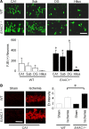
Figure 1.
Figure 1.
Increased ischemia-induced neuronal death and superoxide production in EAAC1−/− mice. A, EAAC1−/− mice show more neuronal death than wild-type (WT) mice after ischemia. Degenerating neurons are identified by Fluoro-Jade B (FJB) staining (green). Confocal fluorescence images show neuronal death in hippocampal CA1, subiculum (Sub), dentate gyrus (DG), and hilus at 3 d after transient ischemia. Scale bar, 100 μm. Bar graph shows quantified cell counts. Data are mean + SEM; n = 5–6, *p < 0.05. B, Ischemia-induced neuronal superoxide production is increased in EAAC1−/− mice. Superoxide is detected by Et fluorescence (red). Images show Et fluorescence in the CA1 hippocampal neurons 3 h after ischemia–reperfusion. Sham-operated mice received surgery without ischemia. Scale bar, 100 μm. Bar graph shows quantified Et fluorescence. n = 4–5, *p < 0.05.
Figure 2.
Figure 2.
Increased ischemia-induced microglial activation in EAAC1−/− mice. Sections were harvested at 3 d after ischemia and immunostained with F4/80 (green). Changes in microglia morphological and F4/80 expression level occur in the hippocampus (CA1 and hilus) and cortex (Ctx). EAAC1−/− mice show increased microglia activation compared with wild-type (WT) mice. Scale bar, 100 μm. Bar graph shows quantified microglia activation in the CA1. Data are mean + SEM; n = 5, *p < 0.05.
Figure 3.
Figure 3.
Basal free zinc and ischemia-induced zinc translocation is elevated in EAAC1−/− mice. A, Images show TSQ zinc fluorescence signals from wild-type (WT) and EAAC1−/− mouse hippocampus. EAAC1−/− mice exhibit an increased TSQ signal in the hilus and stratum radiatum. Scale bar, 500 μm. Graph shows measured TSQ intensity, with BG indicating the background signal measured from the lateral ventricle. B, Higher-magnification images of TSQ-stained hippocampus. Scale bar, 100 μm. The CA1 pyramidal layer shows a brighter signal in EAAC1−/− mice than wild-type mice. Box indicates area of TSQ signal measurement. Graph shows quantified TSQ measurements from CA1. C, Higher-magnification images of TSQ-stained hippocampus from mice 3 d after ischemia shows zinc accumulation in the CA1 pyramidal neurons is greater in EAAC1−/− mice. Higher background signal in the low-power image (A) is attributable to increased image exposure time. Scale bar, 100 μm. Bar graph shows the number of TSQ-positive neurons in CA1 of EAAC1−/− and wild-type mice. Data are mean + SEM; n = 5–6, *p < 0.05.
Figure 4.
Figure 4.
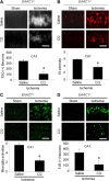
The zinc chelation reduces ischemia-induced zinc translocation, superoxide production, microglia activation, and neuronal death in EAAC1−/− mice. Brains were analyzed at 3 d after ischemia. A, The zinc chelator CQ reduced the number of CA1 TSQ+. Scale bar, 50 μm. B, CQ treatment reduced superoxide production (Et fluorescence) in the hippocampal CA1 area in EAAC1−/− mice. Scale bar, 50 μm. C, CQ treatment reduced microglia activation in the hippocampal CA1 area in EAAC1−/− mice. Scale bar, 50 μm. D, CQ treatment reduced neuron death (FJB+ neurons) in the hippocampal CA1. Scale bar, 100 μm. Data are mean + SEM; n = 5–8, *p < 0.05.
Figure 5.
Figure 5.
Neuronal GSH deficiency in EAAC1−/− mice is reversed by NAC treatment. Reactive thiol content was evaluated in mouse brain using C5 maleimide fluorescence. A, Reduced fluorescence in the neurons of EAAC1−/− hippocampus sections relative to those in the wild-type (WT) slices. This signal was increased in hippocampal sections from mice treated with the cell-permeable cysteine precursor NAC in both wild-type and EAAC1−/− mice. B, Higher-magnification images represent C5 maleimide-stained CA1 pyramidal neurons. Box indicates area of C5 maleimide fluorescence measurement. Bar graph shows the quantified C5 maleimide intensity. Data are mean + SEM; n = 3 in each group, *p < 0.05.
Figure 6.
Figure 6.
NAC reduces vesicular zinc levels and ischemia-induced zinc translocation. A, NAC reduces basal (without ischemia) TSQ intensity in the hippocampal mossy fiber area of both wild-type and EAAC1−/− mice. Scale bar, 500 μm. Data are mean + SEM; n = 3–5, *p < 0.05. B, NAC treatment reduces ischemia-induced zinc translocation in hippocampal CA1. Scale bar, 50 μm. Data are mean + SEM.; n = 5–6, *p < 0.05.
Figure 7.
Figure 7.
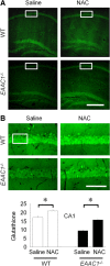
NAC reduces ischemia-induced superoxide production and neuronal death in EAAC1−/− mice. A, NAC treatment reduces Et formation in hippocampal CA1 of both wild-type and EAAC1−/− mice, measured 3 h after ischemia. Scale bar, 50 μm. Data are mean + SEM; n = 3–5, *p < 0.05. B, NAC treatment reduces neuron death (FJB+ neurons) in hippocampal CA1 of both wild-type and EAAC1−/− mice, measured 3 d after ischemia. Scale bar, 50 μm. Data are mean + SEM; n = 5–6, *p < 0.05.
Figure 8.
Figure 8.
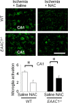
NAC reduces ischemia-induced microglia activation. NAC reduces microglia activation in hippocampal CA1 of both wild-type and EAAC1−/− mice. Scale bar, 50 μm. Data are mean + SEM; n = 5–6, *p < 0.05.
Figure 9.
Figure 9.
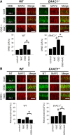
NAC prevents OGD-induced nitrotyrosine production and oxidative injury in EAAC1−/− brain slices. A, Confocal fluorescent images show immunostaining for 4-hydroxynoneal (4HNE, green) and MAP2 (red; identifies the CA1 neuronal perikaria and dendrites) in OGD-treated hippocampal slices. 4HNE formation is higher in EAAC1−/− than wild-type slices (#). 4HNE formation was increased by OGD, and this increase was attenuated by NAC in both wild-type or in EAAC1−/− slices. NAC treatment reduced 4HNE formation (*). Scale bar, 100 μm. AFU, Arbitrary fluorescence units. B, Confocal fluorescent images show immunostaining for nitrotyrosine (NT, green) and MAP2 (red) in OGD-treated hippocampal slices. NT formation is higher in EAAC1−/− than wild-type slices (#). NT formation was increased by OGD, and this increase was attenuated by NAC in both wild-type or in EAAC1−/− slices (*). Scale bar, 100 μm. n = 3, *p < 0.05.

Similar articles

Cited by

References

    1. Aarts M, Iihara K, Wei WL, Xiong ZG, Arundine M, Cerwinski W, MacDonald JF, Tymianski M. A key role for TRPM7 channels in anoxic neuronal death. Cell. 2003;115:863–877. - PubMed
    1. Adlard PA, Cherny RA, Finkelstein DI, Gautier E, Robb E, Cortes M, Volitakis I, Liu X, Smith JP, Perez K, Laughton K, Li QX, Charman SA, Nicolazzo JA, Wilkins S, Deleva K, Lynch T, Kok G, Ritchie CW, Tanzi RE, Cappai R, Masters CL, Barnham KJ, Bush AI. Rapid restoration of cognition in Alzheimer's transgenic mice with 8-hydroxy quinoline analogs is associated with decreased interstitial Abeta. Neuron. 2008;59:43–55. - PubMed
    1. Aizenman E, Stout AK, Hartnett KA, Dineley KE, McLaughlin B, Reynolds IJ. Induction of neuronal apoptosis by thiol oxidation: putative role of intracellular zinc release. J Neurochem. 2000;75:1878–1888. - PubMed
    1. Aoyama K, Suh SW, Hamby AM, Liu J, Chan WY, Chen Y, Swanson RA. Neuronal glutathione deficiency and age-dependent neurodegeneration in the EAAC1 deficient mouse. Nat Neurosci. 2006;9:119–126. - PubMed
    1. Aoyama K, Watabe M, Nakaki T. Regulation of neuronal glutathione synthesis. J Pharmacol Sci. 2008;108:227–238. - PubMed

Publication types