Comparative Study
. 2010 Nov 17;30(46):15654-63.
doi: 10.1523/JNEUROSCI.4340-10.2010.

Long-distance axon regeneration in the mature optic nerve: contributions of oncomodulin, cAMP, and pten gene deletion

Affiliations
Comparative Study

Long-distance axon regeneration in the mature optic nerve: contributions of oncomodulin, cAMP, and pten gene deletion

Takuji Kurimoto et al. J Neurosci. .

Abstract

The inability of retinal ganglion cells (RGCs) to regenerate damaged axons through the optic nerve has dire consequences for victims of traumatic nerve injury and certain neurodegenerative diseases. Several strategies have been shown to induce appreciable regeneration in vivo, but the regrowth of axons through the entire optic nerve and on into the brain remains a major challenge. We show here that the induction of a controlled inflammatory response in the eye, when combined with elevation of intracellular cAMP and deletion of the gene encoding pten (phosphatase and tensin homolog), enables RGCs to regenerate axons the full length of the optic nerve in mature mice; approximately half of these axons cross the chiasm, and a rare subset (∼1%) manages to enter the thalamus. Consistent with our previous findings, the axon-promoting effects of inflammation were shown to require the macrophage-derived growth factor Oncomodulin (Ocm). Elevation of cAMP increased the ability of Ocm to bind to its receptors in the inner retina and augmented inflammation-induced regeneration twofold. Inflammation combined with elevated cAMP and PTEN deletion increased activation of the phosphatidylinositol 3-kinase and mitogen-activated protein kinase signaling pathways and augmented regeneration ∼10-fold over the level induced by either pten deletion or Zymosan alone. Thus, treatments that synergistically alter the intrinsic growth state of RGCs produce unprecedented levels of axon regeneration in the optic nerve, a CNS pathway long believed to be incapable of supporting such growth.


Figures

Figure 1.
Figure 1.
cAMP enhances inflammation-induced axon regeneration in the mouse optic nerve. A–D, Longitudinal sections through the adult mouse optic nerve (ON) showing GAP-43-positive axons distal to the injury site (asterisks) 2 weeks after optic nerve crush. A, Absence of regeneration after surgery alone. B, C, Increased regenerating after intraocular injection of Zymosan (Zymo) (B; 12.5 mg/ml) but not CPT–cAMP alone (C; 50 μm). D, Extensive regeneration induced by Zymosan plus CPT–cAMP. Scale bar, 200 μm. E, Quantitation of axon growth at indicated distances beyond the crush site. ***p < 0.001 compared with optic nerve crush alone. †††p < 0.001 compared with cAMP treatment. #p < 0.05, ###p < 0.001 compared with Zymosan treatment alone. Data in all figures represent group means ± SEM.
Figure 2.
Figure 2.
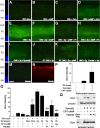
cAMP enhances regeneration in an Oncomodulin-dependent manner. A–D, Longitudinal sections through the adult mouse optic nerve (ON) showing GAP-43-positive axons distal to the injury site (asterisk) 2 weeks after optic nerve crush. All animals received intravitreal injections of Zymosan (Zymo) immediately after optic nerve surgery. Regeneration in the absence of exogenous cAMP is unaffected by the control peptide Pα (A) but is blocked by the Ocm antagonist P1 (B). The enhanced regeneration seen in the presence of exogenous cAMP is unaffected by Pα (C) but is eliminated by P1 (D). Rp-cAMP diminishes Zymosan-induced axonal regeneration (E). Scale bar, 200 μm. F, Quantitation of axon regeneration in the presence or absence of CPT–cAMP, Rp-cAMP, and peptides. †††p < 0.001 compared with Pα injections. #p < 0.05 compared with Zymosan injections (decrease).
Figure 3.
Figure 3.
cAMP regulates Ocm binding in the inner retina. A–N, Sections through the inner retina of mice with optic nerve crush (ONC; A–L) or normal mice (M, N) immunostained to detect Ocm (A–I, M) or βIII tubulin (N) 1 or 7 d after injecting various agents into the posterior chamber of the eye. A–D, Binding of recombinant Ocm in the retina 1 d after optic nerve crush. Ocm is undetectable in the absence of exogenous Ocm or when Ocm or CPT–cAMP are injected alone, but becomes apparent when the latter two are coinjected. E–I, Binding of native Ocm after intraocular injections of Zymosan. Zymosan strongly elevates Ocm immunostaining (E); this is not enhanced by the addition of CPT–cAMP (F). Ocm binding is suppressed by peptide P1 (G) and by Rp-cAMPs (I); the control peptide Pα has a partial effect (H). J, K, Ocm binding 7 d after Zymosan injection is enhanced by CPT–cAMP. L, Reduced immunostaining after preadsorption of the primary antibody with rOcm. M, N, Normal retina shows no detectable Ocm (M). RGCs and their dendrites in the IPL are visualized with an antibody to βIII-tubulin (N). Scale bar: A–N, 25 μm. O, Quantitation of Ocm intensity in the inner plexiform layer of the retina. **p < 0.01 compared with CPT–cAMP alone. †††p < 0.001 compared with Ocm alone. ###p < 0.001 compared with Zymosan plus CPT–cAMP. ON, Optic nerve; Zy, Zymosan. P, Quantitation of Ocm intensity in the inner plexiform layer 7 d after optic nerve (ON) crush. *p < 0.05 compared with Zymosan treatment alone. Q, Ocm binding in the rat retina. One day after injecting Zymosan (Zymo or Z) with or without other agents as indicated, retinas were dissected, homogenized, and fractionated into particulate and soluble fractions, and the proteins were separated by SDS-PAGE. Western blotting used a monoclonal antibody to visualize Ocm and a β-actin antibody to evaluate protein loading. Within the particulate fraction, Ocm levels rose above background after the injection of Zymosan, with or without CPT–cAMP, into the vitreous. P1 but not Pα diminished Ocm binding in the particulate fraction but did not affect levels in the soluble protein fraction. GCL, Ganglion cell layer; INL, inner nuclear layer.
Figure 4.
Figure 4.
Synergistic effects of intraocular inflammation, cAMP, and pten gene deletion. PTEN was deleted in RGCs by injecting AAV2–Cre into the eyes of PTENflx/flx mice 2 weeks before optic nerve surgery (ONC). A–D, Longitudinal sections through the optic nerve showing GAP-43-positive axons 2 weeks after optic nerve injury. PTEN deletion caused appreciable regeneration (A) that was not enhanced by CPT–cAMP (B) and only slightly enhanced by intraocular inflammation (C). The combination of PTEN deletion plus Zymosan (Zymo) plus CPT–cAMP resulted in far greater regeneration than any other treatments (D). E–H, Retinal whole mounts immunostained with TUJ1 to visualize RGCs. E, Normal retina. F, Loss of RGCs 2 weeks after optic nerve crush in PTENflx/flx mouse injected with the control virus (AAV2–GFP). G, H, Preservation of RGCs 2 weeks after optic nerve crush in a PTENflx/flx mouse injected with AAV2–Cre to delete PTEN (G). Survival is not enhanced further by intraocular inflammation plus cAMP (H). Scale bar: A–D, 200 μm; E–H, 50 μm. I, Quantitation of axon regeneration after 2 weeks. *p < 0.05, **p < 0.01, ***p < 0.001 compared with PTEN deletion alone. p < 0.05, ††p < 0.01, †††p < 0.001 compared with PTEN deletion plus Zymosan. #p < 0.05, ##p < 0.01 compared with PTEN deletion plus Zymosan plus CPT–cAMP. J, P1 peptide does not diminish regeneration induced by PTEN deletion. K, Quantitation of RGC survival. PTEN deletion more than tripled the number of RGCs surviving 2 weeks after optic nerve damage compared with PTENflx/flx mice injected with control virus. Survival was not enhanced further by intraocular inflammation and/or CPT–cAMP. ***p < 0.001 compared with nerve-damaged controls.
Figure 5.
Figure 5.
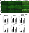
Activation of cell-signaling pathways. A, Sections through the inner retina 3 d after optic nerve crush (ONC) alone or combined with intraocular Zymosan (Zymo) plus CPT–cAMP. PTENflx/flx mice were injected 2 weeks earlier with either AAV2–GFP, allowing RGCs to continue expressing PTEN (PTEN+), or AAV2–Cre to excise the pten gene in RGCs (PTEN−). Sections were stained with antibodies to the phosphorylated (activated) forms of MAP kinase (pMAPK), Akt (pAkt), or S6K (pS6K) as indicated, followed by fluorescent secondary antibodies. Scale bar, 25 μm. GCL, Ganglion cell layer. B, Quantitation of fluorescence in βIII-tubulin-positive RGCs and in the IPL. *p < 0.05, **p < 0.01, ***p < 0.001 compared with PTEN-positive mice with optic nerve crush (ONC) alone. p < 0.05, †††p < 0.001 compared with PTEN-positive mice injected with Zymosan (Zymo) plus CPT–cAMP after optic nerve crush.
Figure 6.
Figure 6.
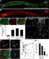
Synergistic interactions lead to long-distance regeneration after 6 weeks. A, B, Regenerating axons extend the full length of the optic nerve in mice lacking the pten gene in RGCs and exposed to intraocular inflammation plus CPT–cAMP. Mice received intraocular injections of CTB 4 d before being prepared for histology. Nerves were double immunostained for GAP-43 (A) and CTB (B). C, D, Enlarged images of the areas enclosed within the white boxes in A and B, respectively. E, Merged image of C and D. F, Quantitation of axon regeneration 3.5 mm from the crush site. The number of lengthy axons increased almost 10-fold between weeks 2 and 6 (***p < 0.001), and most of these axons were double labeled for GAP-43 and CTB. G, Regenerating axons in the optic chiasm. Arrows point to regenerating axons that extend into the thalamus; arrowheads show axons growing into the contralateral optic nerve (ON). H–K, Extension of CTB-labeled axons into the thalamus. Sections were double stained to detect CTB in regenerating axons (red) and NeuN to visualize neurons (green). H and J show double staining, whereas I and K show the axons alone at high contrast. Some axons can be seen in the contralateral optic tract (H, I) and ventral lateral geniculate nucleus (J, K). Arrowheads indicate CTB-labeled axons. L, Schematic drawing through the thalamus showing positions of labeled axons. M, Quantitation of CTB-labeled axons in different segments of the visual pathway. Data are based on four or five cases. dLGN, Dorsal lateral geniculate nucleus; vLGN, ventral lateral geniculate nucleus; OPT, optic tract; Ch, chiasm; CP, cerebral peduncle; Zymo, Zymosan. Scale bars: A, B, 200 μm; C–E, H–K, 50 μm.

References

    1. Bähr M, Wizenmann A. Retinal ganglion cell axons recognize specific guidance cues present in the deafferented adult rat superior colliculus. J Neurosci. 1996;16:5106–5116. - PMC - PubMed
    1. Berry M, Carlile J, Hunter A. Peripheral nerve explants grafted into the vitreous body of the eye promote the regeneration of retinal ganglion cell axons severed in the optic nerve. J Neurocytol. 1996;25:147–170. - PubMed
    1. Berry M, Carlile J, Hunter A, Tsang W, Rosensteil P, Rosustrel P, Sievers J. Optic nerve regeneration after intravitreal peripheral nerve implants: trajectories of axons regrowing through the optic chiasm into the optic tracts. J Neurocytol. 1999;28:721–741. - PubMed
    1. Cai D, Deng K, Mellado W, Lee J, Ratan RR, Filbin MT. Arginase I and polyamines act downstream from cyclic AMP in overcoming inhibition of axonal growth MAG and myelin in vitro. Neuron. 2002;35:711–719. - PubMed
    1. Carracedo A, Pandolfi PP. The PTEN-PI3K pathway: of feedbacks and cross-talks. Oncogene. 2008;27:5527–5541. - PubMed

Publication types

LinkOut - more resources