Comparative Study
. 2010 Nov 17;30(46):15686-94.
doi: 10.1523/JNEUROSCI.4316-10.2010.

Regeneration of new neurons is preserved in aged vomeronasal epithelia

Affiliations
Comparative Study

Regeneration of new neurons is preserved in aged vomeronasal epithelia

Jessica H Brann et al. J Neurosci. .

Abstract

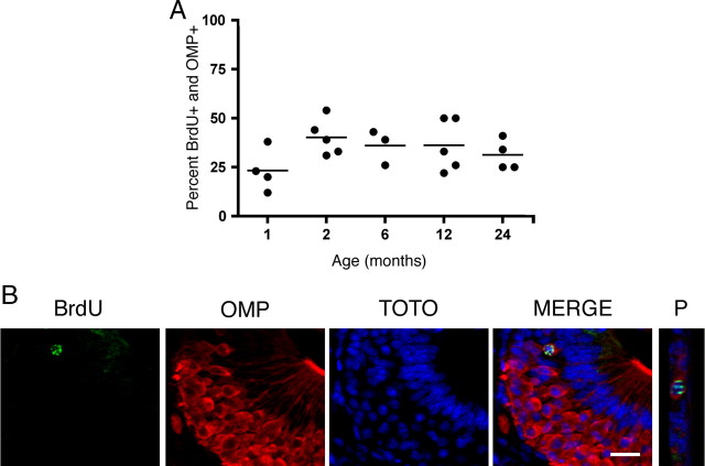
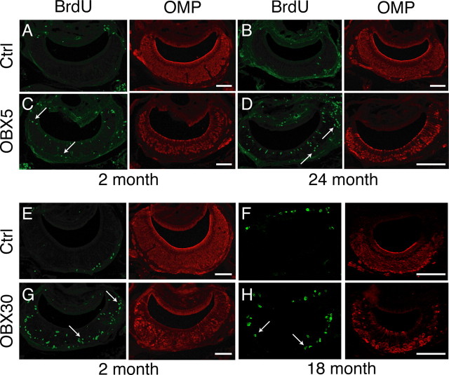
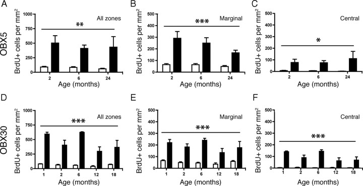
During normal and diseased aging, it is thought the capacity for tissue regeneration and repair in neuronal tissues diminishes. In the peripheral olfactory system, stem cell reservoirs permit regeneration of olfactory and vomeronasal sensory neurons, a unique capacity among neurons. Following injury, a large number of new neurons can be regenerated in a young animal. However, it is unknown whether this capacity for renewal exists in aged proliferative populations. Here, we report that neuronal replacement-associated proliferation continues in the vomeronasal organ of aged (18-24 months) mice. In addition, the potential for the aged stem cell to yield a mature neuron persisted at the same rate as that observed in young animals. Furthermore, the robust regenerative capacity to respond to both acute and sustained injury following olfactory bulbectomy remains intact even in very old animals. Hence, the neuronal epithelium lining the vomeronasal organ is unique in that it contains stem cells capable of generating functional neurons throughout life and in the aged animal in particular. This persistent regenerative capacity provides hope for neuronal replacement therapies in the aged nervous system.


Figures

Figure 1.
Figure 1.
Organization and zones of the vomeronasal organ. A, Schematics depicting the location of the VNO in the sagittal plane (left) and the coronal plane (right). The VNO is a bilaterally symmetrical tubular structure; shown here is one half of a coronal plane as it would appear through the depth of this structure. The marginal zones (M) are found at the extreme dorsal (D) and ventral (V) regions of the VNO. Adjacent to the marginal zones are the intermediate zones (I). In between the two intermediate zones is the central zone (Ce). Adapted with permission from Firestein, 2001 and Mombaerts, 2004. OB, Olfactory bulb; OE, olfactory epithelium; B, blood vessel; C, caudal; L, lumen; R, rostral. B, Incorporation of BrdU (green) in the VNO of a 2-month-old mouse is shown. TOTO-3 is a nuclear marker (blue) and OMP delineates the neuronal area of the VNO (red). Scale bar, 100 μm.
Figure 2.
Figure 2.
Proliferation decreases in the marginal zones but remains constant in the central zone of the VNO with age. A, Incorporation of BrdU (asterisks; green) in the marginal zone of the VNO of a 6-month-old (top) and a 24-month-old (bottom) mouse is shown in the merge of TOTO-3 (nuclear marker; blue) and OMP (red). B, Same as in A but in the central zone of the VNO. C, Quantification of BrdU-positive cells per square millimeter in the marginal zone. *p ≤ 0.0001. D, Quantification of BrdU-positive cells per square millimeter in the central zone. All data were analyzed for statistical significance by one-way ANOVA and SNK pairwise multiple comparison between age groups. n = 5–8 animals per group. Scale bar, 50 μm.
Figure 3.
Figure 3.
Cell death is constant in both the marginal and central zones of the VNO with age. A, Two cells expressing aCasp3 (green), a marker of cell death, in the VNO of a 1-month-old mouse are marked with asterisks. Also shown is OMP (red), TOTO-3 (nuclear marker; blue), and the merge of the three images (right panel). Scale bar, 20 μm. B, C, Quantification of aCasp3-positive cells per square millimeter in the marginal (B) and the central (C) zones revealed no significant difference in the amount of cell death across all age groups. Data were analyzed for statistical significance by one-way ANOVA and SNK pairwise multiple comparison between age groups. p > 0.05; n = 5–8 animals per group. D, The ratio of proliferating cells to dying cells in the marginal zone (filled squares) is significantly different (denoted by asterisks) from that in the central zone (open squares) in young mice (1 month, p < 0.001; 2 month, p < 0.005) but not in aged mice (6–24 months, p > 0.05). The ratio of BrdU-positive to active Caspase-3-positive cells was calculated for each animal in the marginal and central zones and analyzed for statistical significance by a paired Student's t test.
Figure 4.
Figure 4.
Distribution of proliferating cells does not change with age. BrdU (100 mg/kg) was injected 2 h before death. AD, BrdU counts were averaged for each 5% increment along the normalized length of the VNO for each animal in each age group (n = 5–7 per group), with position 0% corresponding to the most rostral extent and position 100% corresponding to the most caudal extent of the VNO. Percentage contribution of each position to the total number of BrdU-positive cells in each animal was calculated. The color represents percentage contribution values as shown in the scale bar, with red corresponding to high percentage contribution and blue corresponding to low percentage contribution. Incorporation of BrdU is highest at the rostral (0%) and caudal (100%) extents of the VNO when data were analyzed either as all zones together (A) or separated by the marginal zone (B), the intermediate zone (C), or the central zone (D). No effect of age was observed in any of the zones. All normalized data were analyzed for statistical significance by two-way ANOVA for the effect of age (not significant) and position (A, B, D, p < 0.0001; C, p < 0.005).
Figure 5.
Figure 5.
Cells incorporating BrdU become mature sensory neurons at the same rate in both young and aged VNO. Mice were injected twice with 50 mg/kg BrdU 2 h apart, killed 30 d following injection, and cells incorporating BrdU were analyzed for the coexpression of OMP, a marker of vomeronasal sensory neuron maturity. A, The percentage of cells incorporating BrdU that reach maturity (OMP-positive) is consistent across all age groups tested (1, 2, 6, 12, and 24 months of age; one-way ANOVA, n = 3–5 animals per group, p > 0.05). A horizontal line represents the mean for each group. B, Representative confocal images of a BrdU-positive cell (green) surrounded by OMP (red). Z-projections of all BrdU-immunoreactive cells were made (projection, P) and examined for overlap with OMP and TOTO-3 (blue, nuclei) to verify that OMP immunoreactivity surrounded a single BrdU-positive nucleus. Scale bar, 20 μm.
Figure 6.
Figure 6.
Acute and sustained unilateral OBX lesion induces increased proliferation in the VNO in young and aged mice. BrdU was administered 2 h before death on d 5 (OBX5; A–D) or d 30 (OBX30; E–H) following OBX. A, BrdU (green) and OMP (red) in nonlesioned control (Ctrl) 2-month-old VNO. B, As in A but for control 24-month-old VNO. C, BrdU and OMP in lesioned (OBX5) 2-month-old VNO. D, As in C but for lesioned 24-month-old VNO. E, BrdU and OMP in nonlesioned control 2-month-old VNO. F, As in E but for control 18-month-old VNO. G, BrdU and OMP in lesioned 2-month-old VNO. H, As in G but for lesioned (OBX30) 18-month-old VNO. Note the increase in BrdU incorporation throughout all zones of the VNO (arrows) in all age groups. Scale bars, 100 μm.
Figure 7.
Figure 7.
Acute and sustained unilateral OBX lesion induces increased proliferation in all zones of the VNO in young and aged mice. AF, BrdU was administered 2 h before death on d 5 (OBX5; A–C) and d 30 (OBX30; D–F) following OBX. A–C, Quantification of BrdU incorporation reveals a significant increase in proliferation in the VNO following acute OBX (black bars) over that in the nonlesion control (white bars) in all zones (A, **p < 0.003), marginal zone (B, ***p < 0.0003), and central zone (C, *p < 0.01), regardless of age (n = 3 per group). D–F, Quantification of BrdU incorporation reveals a significant increase in proliferation in the VNO following sustained OBX (black bars) over that in the nonlesion control (white bars) in all zones (D), marginal zone (E) and central zone (F) (***p < 0.0001) regardless of age (n = 3–7 per group). All data were analyzed by two-way repeated-measures ANOVA for the effects of surgery (p values noted above) and age (not significantly different).

Similar articles

Cited by

References

    1. Ahlenius H, Visan V, Kokaia M, Lindvall O, Kokaia Z. Neural stem and progenitor cells retain their potential for proliferation and differentiation into functional neurons despite lower number in aged brain. J Neurosci. 2009;29:4408–4419. - PMC - PubMed
    1. Bailey KJ, Maslov AY, Pruitt SC. Accumulation of mutations and somatic selection in aging neural stem/progenitor cells. Aging Cell. 2004;3:391–397. - PubMed
    1. Bauer S, Patterson PH. The cell cycle-apoptosis connection revisited in the adult brain. J Cell Biol. 2005;171:641–650. - PMC - PubMed
    1. Caggiano M, Kauer JS, Hunter DD. Globose basal cells are neuronal progenitors in the olfactory epithelium: a lineage analysis using a replication-incompetent retrovirus. Neuron. 1994;13:339–352. - PubMed
    1. Calof AL, Bonnin A, Crocker C, Kawauchi S, Murray RC, Shou J, Wu HH. Progenitor cells of the olfactory receptor neuron lineage. Microsc Res Tech. 2002;58:176–188. - PubMed

Publication types

LinkOut - more resources