. 2011 Feb;89(2):251-7.
doi: 10.1189/jlb.0710395. Epub 2010 Nov 17.

Macrophage motility requires distinct α5β1/FAK and α4β1/paxillin signaling events

Affiliations

Macrophage motility requires distinct α5β1/FAK and α4β1/paxillin signaling events

Michelle Y Abshire et al. J Leukoc Biol. 2011 Feb.

Abstract

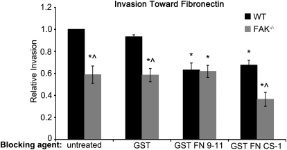
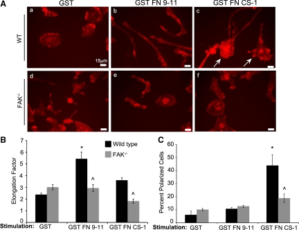
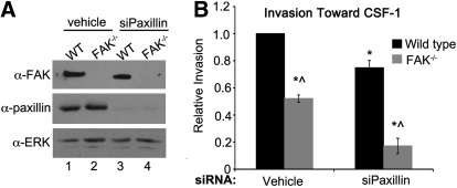
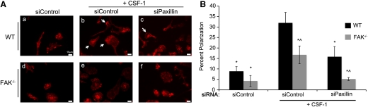
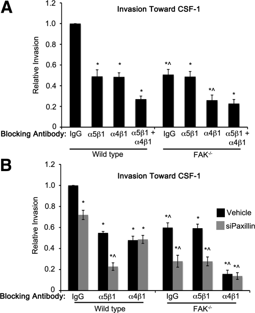
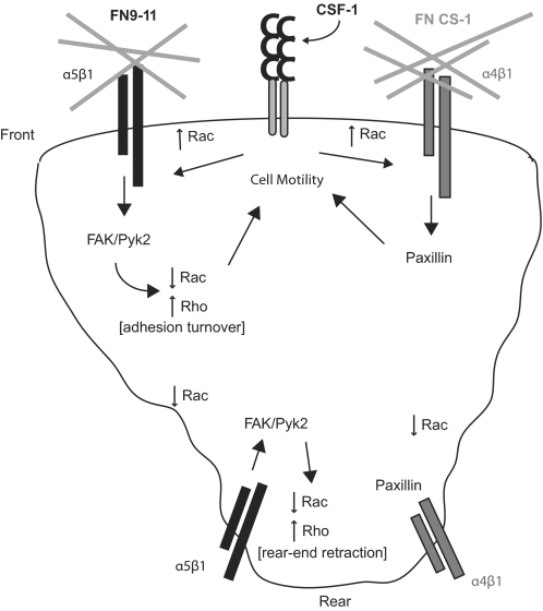
Macrophages function as key inflammatory mediators at sites of infection and tissue damage. Integrin and growth factor receptors facilitate recruitment of monocytes/macrophages to sites of inflammation in response to numerous extracellular stimuli. We have shown recently that FAK plays a role in regulating macrophage chemotaxis and invasion. As FAK is an established downstream mediator of integrin signaling, we sought to define the molecular circuitry involving FAK and the predominant β1 integrin heterodimers expressed in these cells-α4β1 and α5β1. We show that α4β1 and α5β1 integrins are required for efficient haptotactic and chemotactic invasion and that stimulation of these integrin receptors leads to the adoption of distinct morphologies associated with motility. FAK is required downstream of α5β1 for haptotaxis toward FN and chemotaxis toward M-CSF-1 and downstream of α4β1 for the adoption of a polarized phenotype. The scaffolding molecule paxillin functions independently of FAK to promote chemotaxis downstream of α4β1. These studies expand our understanding of β1 integrin signaling networks that regulate motility and invasion in macrophages and thus, provide important new insights into mechanisms by which macrophages perform their diverse functions.


Figures

Figure 1.
Figure 1.. α5β1 and α4β1 control distinct haptotactic invasion pathways in macrophages.
Invasion of WT and FAK−/− BMMs toward FN was measured in matrigel-coated, modified Boyden chambers following pretreatment with the indicated GST proteins. Bars represent the average number of invaded cells divided by the number of WT BMMs that migrated under control (untreated) conditions. *Values significantly different from WT GST treatment conditions; ∧values significantly different from WT cells under the same treatment conditions; n = 8.
Figure 2.
Figure 2.. Stimulation of α4β1 and α5β1 integrins leads to FAK-dependent changes in macrophage morphology.
(A) Representative fields of CSF-1-starved WT and FAK−/− BMMs following 5 min stimulation with the indicated GST proteins. Cells were fixed and stained for phalloidin. Cell elongation (B) and polarization (C) were determined as described in Materials and Methods. *Values significantly different from WT cells treated with GST; ^values significantly different from WT cells under the same treatment conditions; n = 3–4.
Figure 3.
Figure 3.. FAK and paxillin regulate CSF-1-dependent macrophage invasion through separate signaling pathways.
(A) Immunoblot analysis showing paxillin knockdown under control (Lanes 1 and 2) and siPaxillin-treated conditions (lanes 3 and 4) in WT and FAK−/− BMMs. (B) Relative invasion toward CSF-1 of WT and FAK−/− BMMs treated with vehicle or siRNAs targeting paxillin. *Values that are significantly different from vehicle-treated WT cells; ^values that are significantly different from WT cells under the same treatment conditions; n = 6.
Figure 4.
Figure 4.. FAK and paxillin regulate CSF-1-dependent macrophage polarization through separate signaling pathways.
(A) Representative fields of CSF-1-starved WT and FAK−/− BMMs treated with siControl or siPaxillin and stained with phalloidin. (b, c, e, and f). Cells were stimulated with CSF-1 for 6 h. Arrows indicate areas of actin accumulation at the front of polarized cells. (B) Quantification of cell polarization. *Values that are significantly different from WT siControl-treated BMMs stimulated with CSF-1; ^values that are significantly different from WT cells under the same treatment conditions; n = 7.
Figure 5.
Figure 5.. Chemotaxis of macrophages toward CSF-1 requires separate α5β1/FAK and α4β1/paxillin signaling pathways.
(A) Invasion of WT and FAK−/− BMMs toward CSF-1 was measured in the presence of the indicated function-blocking antibodies. Bars represent the average number of migrated cells divided by the number of WT BMMs that migrated under control (IgG-treated) conditions. (B) Invasion was measured as above in control (Vehicle, black bars) or siPaxillin-treated (gray bars) WT and FAK−/− BMMs. *Values significantly different from WT IgG control-treated cells; ^values significantly different from WT cells under the same conditions; n = 3–8.
Figure 6.
Figure 6.. Regulation of macrophage polarity and motility.
We propose a model in which α5β1 couples with FAK and α4β1 with paxillin to regulate macrophage polarity and motility through modulation of Rac and Rho activity.

References

    1. Kummer C., Ginsberg M. H. (2006) New approaches to blockade of α4-integrins, proven therapeutic targets in chronic inflammation. Biochem. Pharmacol. 72, 1460–1468 - PubMed
    1. Rugtveit J., Brandtzaeg P., Halstensen T. S., Fausa O., Scott H. (1994) Increased macrophage subset in inflammatory bowel disease: apparent recruitment from peripheral blood monocytes. Gut 35, 669–674 - PMC - PubMed
    1. Marshall D., Cameron J., Lightwood D., Lawson A. D. (2007) Blockade of colony stimulating factor-1 (CSF-I) leads to inhibition of DSS-induced colitis. Inflamm. Bowel Dis. 13, 219–224 - PubMed
    1. Mantovani A., Sica A. (2010) Macrophages, innate immunity and cancer: balance, tolerance, and diversity. Curr. Opin. Immunol. 22, 231–237 - PubMed
    1. Webb S. E., Pollard J. W., Jones G. E. (1996) Direct observation and quantification of macrophage chemoattraction to the growth factor CSF-1. J. Cell Sci. 109, 793–803 - PubMed

Publication types

MeSH terms