. 2010 Dec 7;107(49):21087-92.
doi: 10.1073/pnas.1011242107. Epub 2010 Nov 17.

Role for a somatically diversified lectin in resistance of an invertebrate to parasite infection

Affiliations

Role for a somatically diversified lectin in resistance of an invertebrate to parasite infection

Patrick C Hanington et al. Proc Natl Acad Sci U S A. .

Abstract

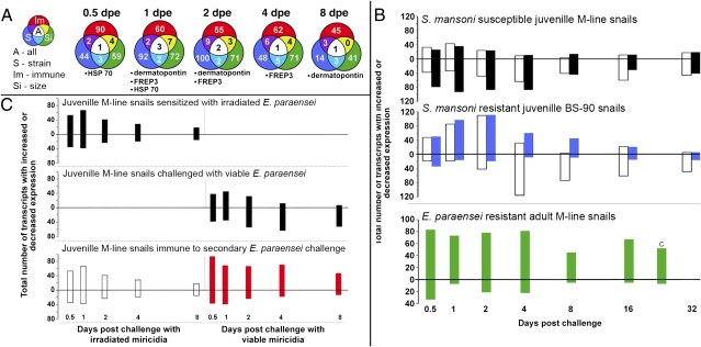
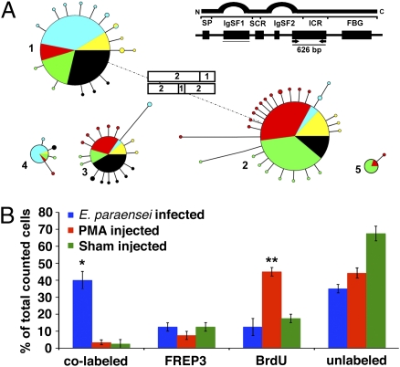
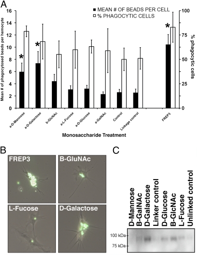
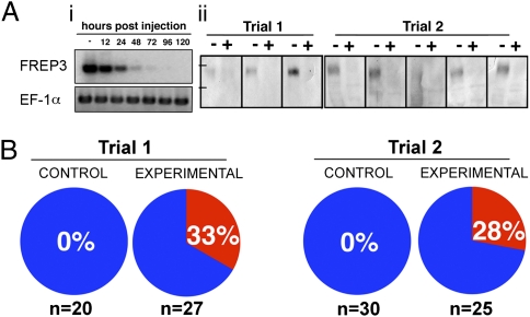
Invertebrates lack adaptive immune systems homologous to those of vertebrates, yet it is becoming increasingly clear that they can produce diversified antigen recognition molecules. We have previously noted that the snail Biomphalaria glabrata produces a secreted lectin, fibrinogen-related protein 3 (FREP3), unusual among invertebrate defense molecules because it is somatically diversified by gene conversion and point mutation. Here we implicate FREP3 in playing a central role in resistance to a major group of snail pathogens, digenetic trematodes. FREP3 is up-regulated in three models of resistance of B. glabrata to infection with Schistosoma mansoni or Echinostoma paraensei, and functions as an opsonin favoring phagocytosis by hemocytes. Knock-down of FREP3 in resistant snails using siRNA-mediated interference resulted in increased susceptibility to E. paraensei, providing a direct link between a gastropod immune molecule and resistance to trematodes. FREP3 up-regulation is also associated with heightened responsiveness following priming with attenuated digenetic trematodes (acquired resistance) in this model invertebrate immune system.


Conflict of interest statement

The authors declare no conflict of interest.

Figures

Fig. 1.
Fig. 1.
Microarray studies of responses of B. glabrata to trematode infection, including three different models of resistance. (A) Venn diagrams showing up-regulated transcripts at different days postexposure (dpe) that are shared among the three forms of resistance to trematode infection. Outer colors of the Venn circles correspond with bar colors shown in A and B. Listed below each time point are the transcripts common to all three resistance models (size, strain, and acquired immune resistance. (B) Enumeration of up-regulated (above zero line) and down-regulated array responses (below zero line) over a time course from 0.5 to 32 d postexposure for juvenile M-line strain snails susceptible to both S. mansoni (open bars) and E. paraensei (black bars); BS-90 strain snails susceptible to E. paraensei (open bars) but resistant to S. mansoni (blue bars); and adult M-line snails resistant to E. paraensei (green bars). In this latter experiment, the transcripts represented on the graph are ones that are up-regulated as a result of size resistance, with those up-regulated during challenge of susceptible small M-line snails subtracted. (C) Summary of transcriptional profiles associated with induction of acquired resistance to E. paraensei in M-line snails: sensitized control snails exposed only to irradiated miracidia, which fail to develop; challenge control snails exposed only to normal (viable) miracidia, which establish successful infections; and experimental snails sensitized with irradiated miracidia and challenged 8 d later with viable miracidia that are killed because of the development of acquired resistance (12). That acquired resistance occurred was verified by exposure of snails to determine if they became infected (shed cercariae): 2 of 59 (3%) snails exposed only to irradiated miracidia, 78 of 84 (93%) exposed only to viable miracidia, and 13 of 94 (14%) exposed first to irradiated miracidia and later challenged with viable miracidia became infected.
Fig. 2.
Fig. 2.
(A) Presence of variant FREP3 genomic sequences in the indicated 626-bp portion of the molecule was ascertained among multiple subsets of 20–40 hemocytes taken from each of five snails (identified by different colors). The five large circles (diameter indicating frequency of recovery) represent the presumptive FREP3 source sequences. Note that the five snails had mostly the same source sequences. The attached small satellite circles (diameter representing recovery rate from 1–3) represent relatively rare FREP3 variants recovered from particular hemocyte subsets that were derived from a source sequence by point mutation (length of connecting lines represents number of differing nucleotides, range 1–6). One variant sequence of source sequence 1 was recovered from two individual snails (satellite circle with two colors). Rectangles indicate additional novel sequences generated by gene conversion, with the contributing source sequences indicated by numbers. The observation of two occurrences does not imply that gene conversion involving FREP3 sequences is rare, as no more than 800 hemocytes were sampled. Our data are consistent with the contribution of gene conversion (additional to point mutations) to extensive somatic diversification of FREP genes as recorded from whole body tissues of B. glabrata (3). (B) Circulating hemocytes taken from snails with 8-d E. paraensei infections were more likely to exhibit both BrdU incorporation (indicative of recent origin) and expression of FREP3 transcripts than hemocytes from snails either injected with PMA or sham injected. *Significant differences from sham-injected controls (P < 0.05, one-way ANOVA). Bars indicate SE.
Fig. 3.
Fig. 3.
FREP3 is involved in detection of monosaccharides and is able to enhance phagocytosis. (A) Streptavidin beads conjugated to different monosaccharides or to purified FREP3 were injected into snails; 2 h later, hemocytes were removed and assayed for presence of beads. Graph shows mean number of beads phagocytosed per hemocyte for 100 hemocytes counted from each snail (n = 10) (filled bars, left axis). Also shown is the percentage of cells counted that had phagocytosed beads (open bars, right axis). *Significant difference from corresponding control (P < 0.05, one-way ANOVA). Bars indicate SE. (B) Hemocytes observed during the phagocytosis experiment with varying numbers of beads within. Beads were conjugated with the substance indicated on each figure. (C) Monosaccharide-conjugated beads were incubated with cell-free snail plasma. Polypeptides were then solubilized from the beads, run on an SDS/PAGE gel, and transferred to nitrocellulose. The Western blot was probed with anti-FREP3 antibody to show which sugars were bound by FREP3.
Fig. 4.
Fig. 4.
RNAi-mediated knock-down of FREP3. (A) Confirmation of transcriptional knock-down (i) using representative results of RT-PCR assays on samples taken from unexposed control snails between 0 and 120 h postinjection. FREP3 knockdown specificity was confirmed using an elongation factor 1-α (EF-1α) endogenous control. Protein level knock-down (ii) shown for two trials, each a Western blot analysis loading 100 μg plasma protein from eight individual snails either before (−) or 4 d after (+) injection of FREP3 specific siRNA. Tick marks represent standard size markers of 75 kDa (Lower) and 100 kDa (Upper). (B) Results of two trials comparing the effects of injection of either GFP oligos (control) or four 27mer FREP3 oligos (experimental). The percentage of snails in each group found to contain rediae at 12–14 dpe is shown and indicated by the red pie segments. The number of snails injected for each group is shown at the bottom of the figure.

Similar articles

Cited by

References

    1. Jones JD, Dangl JL. The plant immune system. Nature. 2006;444:323–329. - PubMed
    1. Alper S, McBride SJ, Lackford B, Freedman JH, Schwartz DA. Specificity and complexity of the Caenorhabditis elegans innate immune response. Mol Cell Biol. 2007;27:5544–5553. - PMC - PubMed
    1. Zhang SM, Adema CM, Kepler TB, Loker ES. Diversification of Ig superfamily genes in an invertebrate. Science. 2004;305:251–254. - PubMed
    1. Watson FL, et al. Extensive diversity of Ig-superfamily proteins in the immune system of insects. Science. 2005;309:1874–1878. - PubMed
    1. Rast JP, Smith LC, Loza-Coll M, Hibino T, Litman GW. Genomic insights into the immune system of the sea urchin. Science. 2006;314:952–956. - PMC - PubMed

Publication types

Associated data