. 2010 Dec 7;107(49):21034-9.
doi: 10.1073/pnas.1007866107. Epub 2010 Nov 17.

Sumoylation activates the transcriptional activity of Pax-6, an important transcription factor for eye and brain development

Affiliations

Sumoylation activates the transcriptional activity of Pax-6, an important transcription factor for eye and brain development

Qin Yan et al. Proc Natl Acad Sci U S A. .

Abstract

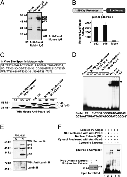
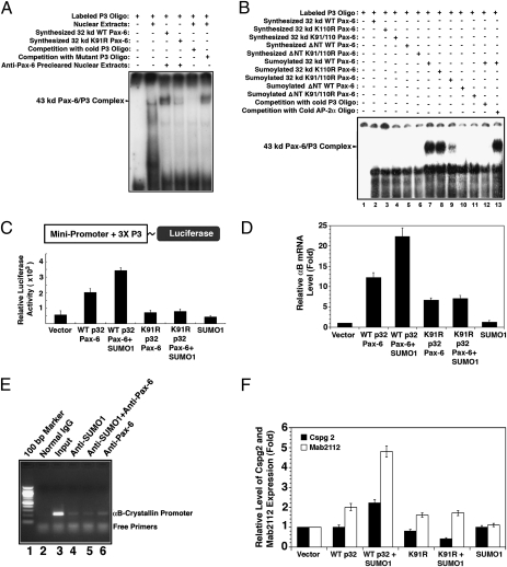
Pax-6 is an evolutionarily conserved transcription factor regulating brain and eye development. Four Pax-6 isoforms have been reported previously. Although the longer Pax-6 isoforms (p46 and p48) bear two DNA-binding domains, the paired domain (PD) and the homeodomain (HD), the shorter Pax-6 isoform p32 contains only the HD for DNA binding. Although a third domain, the proline-, serine- and threonine-enriched activation (PST) domain, in the C termini of all Pax-6 isoforms mediates their transcriptional modulation via phosphorylation, how p32 Pax-6 could regulate target genes remains to be elucidated. In the present study, we show that sumoylation at K91 is required for p32 Pax-6 to bind to a HD-specific site and regulate expression of target genes. First, in vitro-synthesized p32 Pax-6 alone cannot bind the P3 sequence, which contains the HD recognition site, unless it is preincubated with nuclear extracts precleared by anti-Pax-6 but not by anti-small ubiquitin-related modifier 1 (anti-SUMO1) antibody. Second, in vitro-synthesized p32 Pax-6 can be sumoylated by SUMO1, and the sumoylated p32 Pax-6 then can bind to the P3 sequence. Third, Pax-6 and SUMO1 are colocalized in the embryonic optic and lens vesicles and can be coimmunoprecipitated. Finally, SUMO1-conjugated p32 Pax-6 exists in both the nucleus and cytoplasm, and sumoylation significantly enhances the DNA-binding ability of p32 Pax-6 and positively regulates gene expression. Together, our results demonstrate that sumoylation activates p32 Pax-6 in both DNA-binding and transcriptional activities. In addition, our studies demonstrate that p32 and p46 Pax-6 possess differential DNA-binding and regulatory activities.


Conflict of interest statement

The authors declare no conflict of interest.

Figures

Fig. 1.
Fig. 1.
Detection of four Pax-6 isoforms and exploration of DNA-binding and transcriptional activities of p32 and p46 Pax-6. (A) Detection of different Pax-6 isoforms in newborn mouse eyes. Note that p46 and p32 are present in relatively high levels. In contrast, p43 is much reduced. (B) Both p46 and p32 Pax-6 isoforms positively regulate the expression of αB-crystallin as tested in FHL-124 cells. (C) (Upper) In vitro site-specific mutagenesis-generated mutants mimicking constant dephosphorylation (5A) or phosphorylation (5D) in human Pax-6 (WT). (Lower) Western blot analysis of in vitro-generated mutant or wild-type p32 and p46 Pax-6 proteins; note their differential electrophoretic mobilities. In addition, the mutant and wild-type 46-kDa Pax-6 plasmids generate both p46 and p32 because of the activation of an internal ATG initiator. (D) In vitro-synthesized p32 and p46 Pax-6 cannot bind to the P3 sequence. In contrast, nuclear extracts from HLE cells and the P3 sequence form three DNA–protein interacting complexes. The arrowhead designates the complex derived from Pax-6 and the P3 sequence (see legend of Fig. 2 for explanation). (E) Western blot analysis of different Pax-6 isoforms in cytosolic (C) and nuclear (N) extracts of FHL-124 cells. (F) EMSA demonstrating that p43 Pax-6 from FHL-124 cytosol and the P3 sequence form a strong interacting complex. The relative amounts of nuclear and cytosolic p43 Pax-6 are shown in the Western blot.
Fig. 2.
Fig. 2.
Demonstration that sumoylation of the p32 Pax-6 activates its DNA-binding activity. (A) Gel mobility-shifting assay showing that in vitro-translated p32 Pax-6 can bind to the P3 sequence only after preincubation with Pax-6–depleted FHL-124 nuclear extract. (B) Western blot analysis indicating the amounts of in vitro-synthesized mutant and wild-type p32 Pax-6 used for EMSA described in C. (C) EMSA demonstrating that phosphorylation and dephosphorylation modulate DNA binding of p32 Pax-6 to the P3 sequence. (D) EMSA demonstrating that p32 but not p46 Pax-6 binds to the P3 sequence. (E) Western blot analysis of in vitro-synthesized p46 and p32 proteins from Pax-6 full-length cDNA without (lane 1) or with preclearance by serum 11 (lane 2) or by serum 14 (lane 3). (F) EMSA demonstrating that sumoylation is necessary for p32 Pax-6 to bind to the P3 sequence. (G) Demonstration that p32 Pax-6 can be sumoylated by SUMO1 in vitro. (a) The in vitro-generated GST–p32 Pax-6 fusion protein was purified by thrombin cleavage and GST column. (b and c) Purified p32 Pax-6 was subjected to sumoylation with a kit from Biomol (UW8955). After sumoylation, the reaction products were identified by anti-SUMO1 (b) or anti–Pax-6 antibodies (c). Arrowheads indicate p43 Pax-6. (H) EMSA demonstrating that sumoylation of p32 Pax-6 in vitro activates its DNA binding to the P3 sequence.
Fig. 3.
Fig. 3.
Demonstration that sumoylation of p32 Pax-6 at K91 activates its DNA-binding and transcriptional activities. (A) EMSA showing that sumoylation at K91 activates p32 Pax-6 DNA-binding activity. (B) EMSA showing that deletion of the N terminus of p32 Pax-6 abolishes its DNA-binding activity (lanes 5 and 10). In contrast, mutation in the other putative sumoylation site, K110, has no effect on the DNA binding of p32 Pax-6 (lane 8). (C) Sumoylation of p32 Pax-6 activates its transactivity on the luciferase reporter gene as tested in FHL-124 cells. (D) Sumoylation of p32 Pax-6 up-regulates expression of the endogenous αB-crystallin gene in ARPE cells. ARPE-19 cells were transfected with vector alone or with wild-type or K91R p32 Pax-6 with or without SUMO1 as indicated. After 36 h, the transfected cells were harvested for preparation of total RNAs, which were used for quantitative real-time PCR as described in Methods. Note that αB-crystallin mRNA from vector-transfected cells is considered as 1.0. Transfection of wild-type p32 Pax-6 yielded a 12-fold increase in αB-crystallin mRNA expression. Cotransfection of the wild-type p32 Pax-6 with SUMO1 leads to an additional 11-fold enhancement of the αB-crystallin mRNA expression. The K91R mutant substantially decreased its transactivity. (E) ChIP assay demonstrating that Pax-6 sumoylated by SUMO1 binds directly to the αB-crystallin gene promoter in ARPE-19 cells. Lane 5 indicates ChIP result from sequential precipitations, first by anti-SUMO1 and then by anti–Pax-6 antibodies. (F) Sumoylation of p32 Pax-6 up-regulates expression of the endogenous Cspg2 and Mab2112 genes in αTN4-1 cells. αTN4-1 cells were transfected and processed as described in Fig. 3D. Cotransfection of the wild-type p32 Pax-6 with SUMO1 leads to a 2.2-fold enhancement of Cspg2 mRNA expression and a 5.1-fold increase of Mab2112 mRNA expression in αTN4-1 cells. The p32 K91R mutant substantially decreased its transactivity.
Fig. 4.
Fig. 4.
Presence of p32 Pax-6 sumoylation in the developing mouse embryonic eye. (A and B) Immunohistochemistry analysis of Pax-6 and SUMO1 in the optic vesicle and lens vesicle of a mouse embryonic eye. Note that both Pax-6 and SUMO1 are clearly detected in the optical vesicle at ED 9.5 (A) and in the lens vesicle and retina tissues at ED 11.5 (B). In most cases, Pax-6 and SUMO1 overlap each other to generate the yellow fluorescence (arrowheads) (C and D) Total proteins were extracted from ED 11.5 embryonic eyes and were used for immunoprecipitation-linked Western blot analysis. Note that p43 Pax-6 (arrowhead in C) immunoprecipitated by anti-SUMO1 can be detected by anti–Pax-6 antibody. Similarly, p43 Pax-6 (arrowhead in D) immunoprecipitated by anti–Pax-6 can be detected by anti-SUMO1 antibody. L, lens; OV, optic vesicle; R, retina.

Similar articles

Cited by

References

    1. Mansouri A, Hallonet M, Gruss P. Pax genes and their roles in cell differentiation and development. Curr Opin Cell Biol. 1996;8:851–857. - PubMed
    1. Callaerts P, Halder G, Gehring WJ. PAX-6 in development and evolution. Annu Rev Neurosci. 1997;20:483–532. - PubMed
    1. Ton CC, et al. Positional cloning and characterization of a paired box- and homeobox-containing gene from the aniridia region. Cell. 1991;67:1059–1074. - PubMed
    1. Glaser T, Walton DS, Maas RL. Genomic structure, evolutionary conservation and aniridia mutations in the human PAX6 gene. Nat Genet. 1992;2:232–239. - PubMed
    1. Wilson D, Sheng G, Lecuit T, Dostatni N, Desplan C. Cooperative dimerization of paired class homeo domains on DNA. Genes Dev. 1993;7:2120–2134. - PubMed

Publication types