. 2011 Feb;300(2):C318-27.
doi: 10.1152/ajpcell.00172.2010. Epub 2010 Nov 17.

Intracellular signaling pathways regulating net protein balance following diaphragm muscle denervation

Affiliations

Intracellular signaling pathways regulating net protein balance following diaphragm muscle denervation

Heather M Argadine et al. Am J Physiol Cell Physiol. 2011 Feb.

Abstract

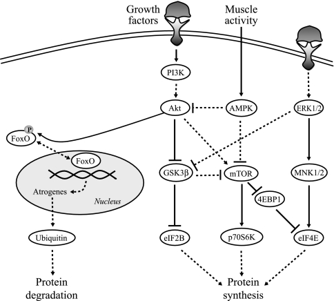
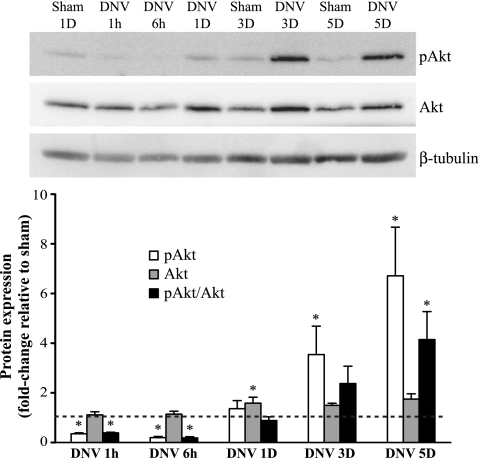
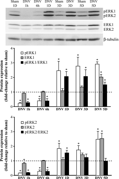
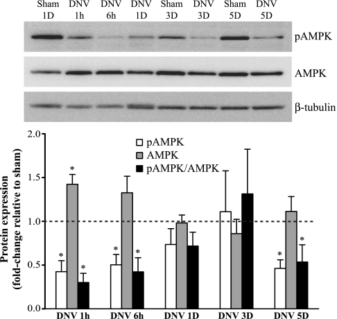
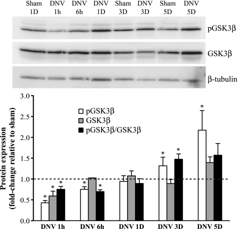
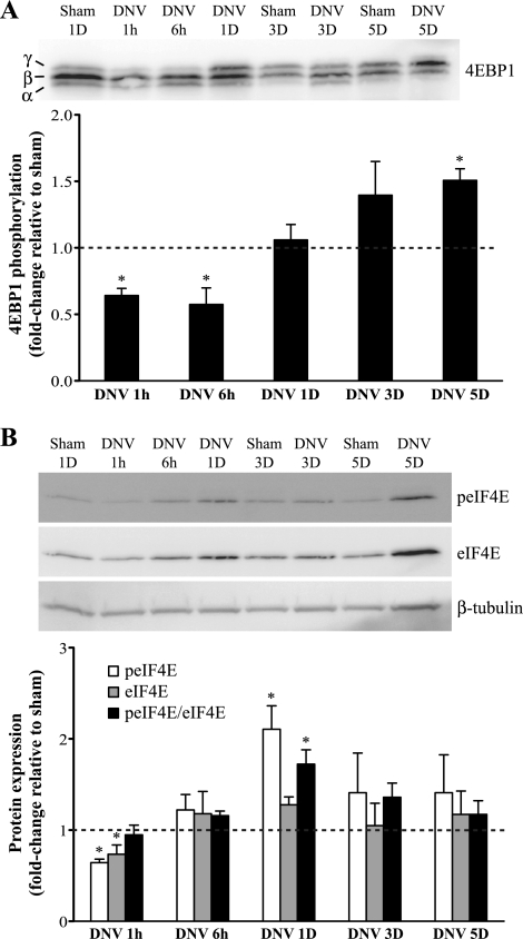
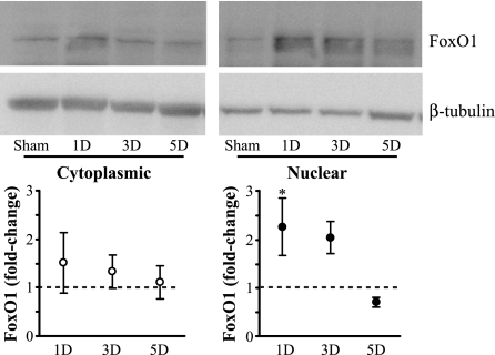
Unilateral denervation (DNV) of rat diaphragm muscle increases protein synthesis at 3 days after DNV (DNV-3D) and degradation at DNV-5D, such that net protein breakdown is evident by DNV-5D. On the basis of existing models of protein balance, we examined DNV-induced changes in Akt, AMP-activated protein kinase (AMPK), and ERK&frac12; activation, which can lead to increased protein synthesis via mammalian target of rapamycin (mTOR)/p70S6 kinase (p70S6K), glycogen synthase kinase-3β (GSK3β), or eukaryotic initiation factor 4E (eIF4E), and increased protein degradation via forkhead box protein O (FoxO). Protein phosphorylation was measured using Western analyses through DNV-5D. Akt phosphorylation decreased at 1 h and 6 h after DNV compared with sham despite decreased AMPK phosphorylation. Both Akt and AMPK phosphorylation returned to sham levels by DNV-1D. Phosphorylation of their downstream effector mTOR (Ser2481) did not change at any time point after DNV, and phosphorylated p70S6K and eIF4E-binding protein 1 (4EBP1) increased only by DNV-5D. In contrast, ERK&frac12; phosphorylation and its downstream effector eIF4E increased 1.7-fold at DNV-1D and phosphorylated GSK3β increased 1.5-fold at DNV-3D (P < 0.05 for both comparisons). Thus, following DNV there are differential effects on protein synthetic pathways with preferential activation of GSK3β and eIF4E over p70S6K. FoxO1 nuclear translocation occurred by DNV-1D, consistent with its role in increasing expression of atrogenes necessary for subsequent ubiquitin-proteasome activation evident by DNV-5D. On the basis of our results, increased protein synthesis following DNV is associated with changes in ERK&frac12;-dependent pathways, but protein degradation results from downregulation of Akt and nuclear translocation of FoxO1. No single trigger is responsible for protein balance following DNV. Protein balance in skeletal muscle depends on multiple synthetic/degradation pathways that should be studied in concert.


Figures

Fig. 1.
Fig. 1.
Simplified model for the intersection of signaling pathways regulating protein synthesis and degradation. Arrows indicate activating events, whereas perpendicular lines indicate inhibitory events. The solid lines represent direct activation. The dashed lines represent indirect activation, whereby intermediate steps are involved but are not specified in this schematic. Protein synthesis is regulated by protein kinase B (Akt), p44/42 MAPK (ERK½), and AMP-activated protein kinase (AMPK), leading to activation of the downstream targets mammalian target of rapamycin (mTOR), glycogen synthase kinase-3β (GSK3β), MAPK-interacting kinases ½ (MNK½), p70S6 kinase (p70S6K), eIF4E-binding protein 1 (4EBP1), and eukaryotic initiation factors 2B and 4E (eIF2B and eIF4E). Conversely, Akt is responsible for the phosphorylation status of forkhead box protein (FoxO). If FoxO is phosphorylated by Akt, it leaves the nucleus and becomes inactive, thus preventing protein degradation. If Akt activity is suppressed, FoxO becomes dephosphorylated, translocates to the nucleus, and exerts its transcriptional effects on atrogenes to induce protein degradation through the ubiquitin/proteasome pathway. PI3K, phosphoinositide 3-kinase.
Fig. 2.
Fig. 2.
Unilateral denervation (DNV)-induced changes in phosphorylation of Akt, as detected by Western analyses. Top: representative immunoblot of phospho-Akt, Akt, and β-tubulin for each DNV time point [in hours (h) or days (D)]. Bottom: relative expression (means ± SE) of phospho-Akt, Akt, and the ratio of phospho-Akt to total Akt, compared with sham control after normalization to β-tubulin. *Significantly different (P < 0.05; n = 6 animals per time point) from the average of all sham controls.
Fig. 3.
Fig. 3.
DNV-induced changes in phosphorylation of ERK½, as detected by Western analyses. Top: representative immunoblot of phospho-ERK½, ERK½, and β-tubulin for each DNV time point. Bottom: relative expression (means ± SE) of phospho-ERK½, ERK½, and the ratio of phospho-ERK½ to total ERK½, compared with sham control after normalization to β-tubulin. *Significantly different (P < 0.05; n = 6) from the average of all sham controls.
Fig. 4.
Fig. 4.
DNV-induced changes in phosphorylation of AMPK, as detected by Western analyses. Top: representative immunoblot of phospho-AMPK, AMPK, and β-tubulin for each DNV time point. Bottom: relative expression (means ± SE) of phospho-AMPK, AMPK, and the ratio of phospho-AMPK to total AMPK, compared with sham control after normalization to β-tubulin. *Significantly different (P < 0.05; n = 6) from the average of all sham controls.
Fig. 5.
Fig. 5.
DNV-induced changes in phosphorylation of mTOR (A) and p70S6K (B), as detected by Western analyses. A, top: representative immunoblot of phospho-mTOR, mTOR, and β-tubulin for each DNV time point. Bottom: relative expression (means ± SE) of phospho-mTOR, mTOR, and the ratio of phospho-mTOR to total mTOR, compared with sham control after normalization to β-tubulin. *Significantly different (P < 0.05; n = 5) from the average of all sham controls. B, top: representative immunoblot of phospho-p70S6K, p70S6K, and β-tubulin for each DNV time point. Bottom: relative expression (means ± SE) of phospho-p70S6K and p70S6K, compared with sham control after normalization to β-tubulin. *Significantly different (P < 0.05; n = 4) from the average of all sham controls.
Fig. 6.
Fig. 6.
DNV-induced changes in phosphorylation of GSK3β, as detected by Western analyses. Top: representative immunoblot of phospho-GSK3β, GSK3β, and β-tubulin for each DNV time point. Bottom: relative expression (means ± SE) of phospho-GSK3β, GSK3β, and the ratio of phospho-GSK3β to total GSK3β, compared with sham control after normalization to β-tubulin. *Significantly different (P < 0.05; n = 6) from the average of all sham controls.
Fig. 7.
Fig. 7.
DNV-induced changes in phosphorylation of 4EBP1 (A) and eIF4E (B), as detected by Western analyses. A, top: representative immunoblot of 4EBP1 for each DNV time point. Bottom: relative expression (means ± SE) of 4EBP1 phosphorylation, calculated by the ratio of the hyperphosphorylated γ-band relative to the sum of all bands (γ + β + α). *Significantly different (P < 0.05; n = 6) from the average of all sham controls. B, top: representative immunoblot of phospho-eIF4E, eIF4E, and β-tubulin for each DNV time point. Bottom: relative expression (means ± SE) of phospho-eIF4E, eIF4E, and the ratio of phospho-eIF4E to eIF4E, compared with sham control after normalization to β-tubulin. *Significantly different (P < 0.05; n = 5) from the average of all sham controls.
Fig. 8.
Fig. 8.
DNV-induced changes in FoxO1 protein expression in cytoplasmic and nuclear cell fractions, as detected by Western analyses. Top: representative immunoblot of cytoplasmic and nuclear FoxO1 in sham control and at 1, 3, and 5 days after DNV. Bottom: relative expression (means ± SE) of cytoplasmic FoxO1 (left) and nuclear FoxO1 protein expression (right), each compared with sham control for the same cell fraction after normalization to β-tubulin. *Significantly different (P < 0.05; n = 3) from the sham control.

References

    1. Akoumianaki T, Kardassis D, Polioudaki H, Georgatos SD, Theodoropoulos PA. Nucleocytoplasmic shuttling of soluble tubulin in mammalian cells. J Cell Sci 122: 1111–1118, 2009 - PubMed
    1. Anthony JC, Yoshizawa F, Anthony TG, Vary TC, Jefferson LS, Kimball SR. Leucine stimulates translation initiation in skeletal muscle of postabsorptive rats via a rapamycin-sensitive pathway. J Nutr 130: 2413–2419, 2000 - PubMed
    1. Argadine HM, Hellyer NJ, Mantilla CB, Zhan WZ, Sieck GC. The effect of denervation on protein synthesis and degradation in adult rat diaphragm muscle. J Appl Physiol 107: 438–444, 2009 - PMC - PubMed
    1. Baar K, Esser K. Phosphorylation of p70S6k correlates with increased skeletal muscle mass following resistance exercise. Am J Physiol Cell Physiol 276: C120–C127, 1999 - PubMed
    1. Bamman MM, Shipp JR, Jiang J, Gower BA, Hunter GR, Goodman A, McLafferty CL, Jr, Urban RJ. Mechanical load increases muscle IGF-I and androgen receptor mRNA concentrations in humans. Am J Physiol Endocrinol Metab 280: E383–E390, 2001 - PubMed

Publication types

MeSH terms

Substances