. 2010 Dec;120(12):4546-57.
doi: 10.1172/JCI43127. Epub 2010 Nov 15.

Tim-3 expression on PD-1+ HCV-specific human CTLs is associated with viral persistence, and its blockade restores hepatocyte-directed in vitro cytotoxicity

Affiliations

Tim-3 expression on PD-1+ HCV-specific human CTLs is associated with viral persistence, and its blockade restores hepatocyte-directed in vitro cytotoxicity

Rachel H McMahan et al. J Clin Invest. 2010 Dec.

Erratum in

  • J Clin Invest. 2011 Feb 1;121(2):821

Abstract

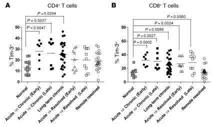
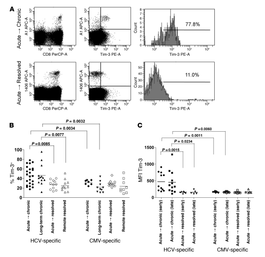
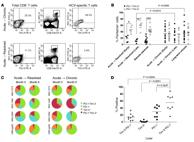
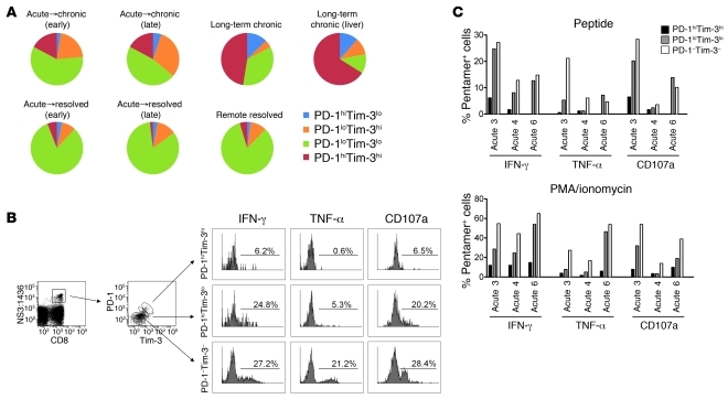
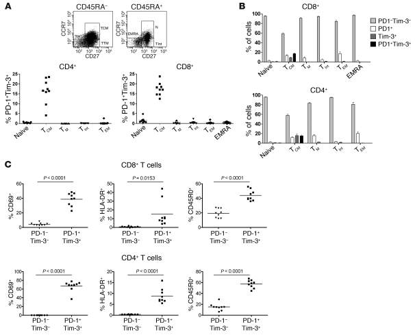
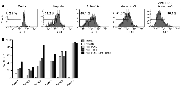
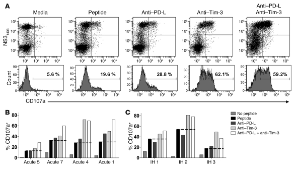
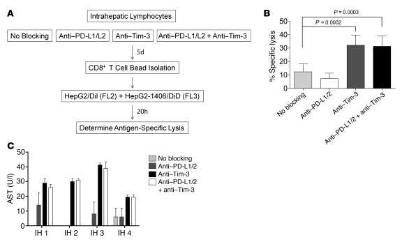
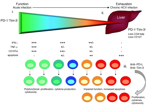
Having successfully developed mechanisms to evade immune clearance, hepatitis C virus (HCV) establishes persistent infection in approximately 75%-80% of patients. In these individuals, the function of HCV-specific CD8+ T cells is impaired by ligation of inhibitory receptors, the repertoire of which has expanded considerably in the past few years. We hypothesized that the coexpression of the negative regulatory receptors T cell immunoglobulin and mucin domain-containing molecule 3 (Tim-3) and programmed death 1 (PD-1) in HCV infection would identify patients at risk of developing viral persistence during and after acute HCV infection. The frequency of PD-1-Tim-3- HCV-specific CTLs greatly outnumbered PD-1+Tim-3+ CTLs in patients with acute resolving infection. Moreover, the population of PD-1+Tim-3+ T cells was enriched for within the central memory T cell subset and within the liver. Blockade of either PD-1 or Tim-3 enhanced in vitro proliferation of HCV-specific CTLs to a similar extent, whereas cytotoxicity against a hepatocyte cell line that expressed cognate HCV epitopes was increased exclusively by Tim-3 blockade. These results indicate that the coexpression of these inhibitory molecules tracks with defective T cell responses and that anatomical differences might account for lack of immune control of persistent pathogens, which suggests their manipulation may represent a rational target for novel immunotherapeutic approaches.


Figures

Figure 1
Figure 1. Differential Tim-3 expression on total CD4+ and CD8+ T cells in acute, chronic, and resolved HCV infection.
T cell populations from acutely infected patients (n = 16) early (<3 months) and late (>6 months) after infection as well as long-term chronic (n = 27), remotely resolved (n = 23), and normal control (n = 10) subjects were identified by staining PBMCs with antibodies against CD3, CD4, and CD8. (A and B) Percent CD4+ and CD8+ T cells expressing Tim-3. Horizontal bars denote means. (A) CD4+ T cells from acute→chronic patients had significantly more Tim-3+ T cells at both early and late time points after infection compared with acute→resolved patients. (B) CD8+ T cells from acutely infected patients had significantly higher Tim-3 levels regardless of viral outcome, but Tim-3 levels returned to normal in remote resolved patients. There was no statistical difference between acute→chronic and acute→resolved patients at comparable phases of infection.
Figure 2
Figure 2. Tim-3 expression on HCV-specific CTLs is higher in acute→chronic patients.
HCV-specific T cell populations were identified by staining with antibodies against CD3, CD8, and a panel of HCV-specific MHC class I pentamers (HLA-A21406, HLA-A22594, and HLA-A11436) along with HLA-A2–rescricted CMV (pp65) pentamer. (A) Representative plots of Tim-3 expression on HCV-specific T cells from 1 acute→chronic and 1 acute→resolved patient. (B and C) Percent pentamer-positive cells expressing Tim-3 (B) and MFI of Tim-3 expression (C) from acute patients (34 responses from 14 patients at 2 time points), long-term chronic patients (13 responses from 11 patients), and remote resolved patients (11 responses from 11 patients). Horizontal bars denote means. Antigen-specific T cells from acute→chronic patients expressed significantly more Tim-3 than did those of acute→resolved patients. A similar expression pattern was seen when comparing long-term chronic with remote resolved patients. Expression of Tim-3 was higher, by both percentage and MFI, on HCV-specific T cells than on CMV-specific T cells in both acute and long-term chronic patients. There was no significant difference in the number of CMV-specific T cells expressing Tim-3 according to HCV outcome.
Figure 3
Figure 3. Dual expression of Tim-3 and PD-1 on virus-specific CTLs predicts acute development of persistence.
HCV-specific T cell populations were identified by staining with antibodies against CD3, CD8, and a panel of MHC class I pentamers (HLA-A21406, HLA-A22594, HLA-A21073, and HLA-A11436). (A) Representative plots showing dual expression of PD-1 and Tim-3 on total CD8+ T cells and HCV-specific T cells from an acute→chronic and an acute→resolved patient. (B) Percentage PD-1+Tim-3+ or PD-1Tim-3 pentamer-positive cells from early and late acute→chronic and acute→resolved, long-term chronic, and remote resolved patients. Antigen-specific T cells from acute→resolved and remote resolved patients had significantly more PD-1+Tim-3+ than PD-1Tim-3 cells (*P = 0.039, **P = 0.0062, ***P = 0.0009). Conversely, the frequencies of PD-1+Tim-3+ and PD-1Tim-3 CTLs were equivalent in acute→chronic patients. (C) Boolean graphs from 2 representative acutely infected patients showing the percentage of HCV- and CMV-specific T cells that expressed Tim-3 and PD-1 at early and late time points after infection. (D) Percent intrahepatic HCV-specific CTLs expressing Tim-3, PD-1, or both. The predominant phenotype of liver-resident CTLs was PD-1+Tim-3+, and the least frequent was PD-1Tim-3+. Horizontal bars in B and D denote means.
Figure 4
Figure 4. PD-1hiTim-3hi CTLs are associated with diminished antiviral cytokine production and chronic infection.
(A) PD-1hiTim-3lo, PD-1loTim-3hi, PD-1loTim-3lo, and PD-1hiTim-3hi pentamer-positive T cells from acute→chronic, acute→resolved, long-term chronic, and remote resolved patients as well as intrahepatic T cells from long-term chronic (liver) patients. The differences between long-term chronic and remote resolved patients were highly statistically significant (see Results). (B and C) PBMCs from acute→chronic patients were stimulated with the HLA-A1–restricted NS31436 epitope or PMA and ionomycin for 5 hours in the presence of the CD107a antibody. Cell surface staining for CD3, CD8, HLA-A11436 pentamer, Tim-3, and PD-1 was carried out, followed by intracellular cytokine staining for IFN-γ and TNF-α. Flow cytometric analysis was used to determine the proportions of the PD-1hiTim-3hi, PD-1loTim-3lo, and PD-1Tim-3 cells producing IFN-γ, TNF-α, and CD107a after stimulation. (B) Histograms from a representative patient with high pentamer frequency (2.2%). (C) Percent cells producing IFN-γ, TNF-α, and CD107a according to PD-1 and Tim-3 expression following stimulation with the NS31436 peptide or PMA and ionomycin. Pentamer frequencies for patients acute 4 and acute 6 are shown in Supplemental Figure 3.
Figure 5
Figure 5. T cells coexpressing Tim-3 and PD-1 display a TCM phenotype.
(A and B) CD4+ and CD8+ T cells from acutely infected patients (n = 9) were stained with antibodies against CD45RA, CCR7, and CD27 to determine differentiation phenotype (see Results). (A) Percent PD-1+Tim-3+ cells within each population. Cells expressing both PD-1 and Tim-3 were predominantly of the TCM phenotype. (B) Percent (± SEM) PD-1Tim-3, PD-1+Tim-3, PD-1Tim-3+, and PD-1+Tim-3+ cells within each population. There were no significant differences in the phenotype of PD-1+Tim-3+ T cells from normal subjects and HCV patients (not shown). (C) CD4+ and CD8+ T cells were stained with antibodies against PD-1, Tim-3, CD69, HLA-DR, and CD45RO, and the PD-1+Tim-3+ and PD-1Tim-3 phenotypes were compared. Horizontal bars denote means. A significant portion of PD-1+Tim-3+ T cells expressed all 3 activation/memory markers.
Figure 6
Figure 6. Blocking Tim-3 and PD-1 enhances T cell proliferation.
PBMCs from 5 acute→chronic patients (acute 2–acute 6) and 1 acute→resolved patient (acute 1; ref. 8) were stained with CFSE and stimulated for 7 days with HLA-A2–restricted NS31406 or HLA-A1–restricted NS31436 peptide, IL-2 (0.5 ng/ml), anti–Tim-3 antibody (10 μg/ml), anti–PD-L1/PD-L2 antibodies (10 μg/ml), or both anti–Tim-3 and anti–PD-L1/PD-L2. HCV-specific T cells were identified by staining with anti-CD8 and HLA-A21406 or HLA-A11436 pentamers and analyzed by flow cytometry for proliferation, as measured by CFSElo cells. (A) Representative histograms from 1 patient showing an increase in CFSElo T cells after dual blocking of Tim-3 and PD-1. Percent HCV-specific T cells that proliferated are shown. (B) Percent CFSElo cells from 6 patients following treatment with the indicated blocking antibodies. Blocking with either PD-1 or Tim-3 alone improved proliferation in 4 of the 6 patients, and dual blocking further enhanced proliferation.
Figure 7
Figure 7. Dual blocking of PD-1 and Tim-3 enhances CD107a expression by HCV-specific T cells.
PBMCs from patients acute 1, acute 4, acute 5, and acute 7 or intrahepatic lymphocytes from long-term chronic (liver) patients (IH 1–IH 3) were stimulated for 7 days with NS31406 or NS31436 peptide (10 μg/ml), IL-2 (0.5 ng/ml), and control IgG, anti–Tim-3, anti–PD-L1/PD-L2, or both anti–Tim-3 and anti–PD-L1/PD-L2 antibodies. Cells were restimulated with peptide, and CD107a antibody and brefeldin A were added to the cultures 5 hours prior to staining with anti-CD8 and HLA-A21406 or HLA-A11436 pentamers. CD107a expression on HCV-specific CTLs was determined by flow cytometric analysis. (A) Representative histograms from 1 acute patient showing an increase in CD107a+ CTLs after dual blocking of Tim-3 and PD-1. (B and C) Percent HCV-specific T cells expressing CD107a from (B) acute→chronic patients or (C) intrahepatic lymphocytes from long-term chronic (liver) patients. Blocking Tim-3 alone or Tim-3 and PD-1 increased CTL cytotoxicity in both PBMCs and intrahepatic lymphocytes. Dotted lines represent CD107a expression after stimulation with peptide alone.
Figure 8
Figure 8. Tim-3 blockade enhances antigen-specific CTL killing of hepatocytes.
(A) Experimental design to assess how Tim-3 and/or PD-1 blockade affects cytotoxicity of HCV-specific CTLs against a hepatocyte cell line expressing an NS3 epitope. Cytotoxicity of NS31406-specific T cells against HepG2 cells transduced with a NS31406–1415 minigene was determined using a variation of the VITAL assay. (B) Intrahepatic lymphocytes were stimulated for 5 days with NS31406 peptide and the indicated blocking antibodies, followed by CD8+ T cell bead isolation. HCV-specific T cells were then cocultured with HepG2 cells expressing the NS31406 minigene at a 0.5:1 effector/target ratio. Percent specific lysis was calculated (see Methods). Values denote mean ± SEM of 6 patients; P values were determined using paired 2-tailed t test. (C) AST levels in the supernatants from B demonstrated an increase in cultures containing the Tim-3–blocking antibody. Values denote mean ± SEM of triplicate samples.
Figure 9
Figure 9. Gradient of function and exhaustion in HCV-specific T cells according to expression of Tim-3 and PD-1.
PD-1Tim-3 T cells (i) demonstrate a highly polyfunctional profile, with production of antiviral cytokines, cytotoxicity, and phenotype consistent with TEM. Higher frequencies of these cells in acute HCV infection correspond to spontaneous viral eradication. At the other end of the spectrum, PD-1+Tim-3+ T cells (ii) accumulate in early HCV infection prior to development of persistence, demonstrate impaired CD4 help (decreased IL-2 production), impaired ability to secrete antiviral cytokines, and a phenotypic profile consistent with TCM. Initially, TNF-α production is impaired, even though CD8+ T cells maintain the ability to produce IFN-γ (ref. and Figure 4B). The liver contains high expression of the ligands for PD-1 and Tim-3 (PD-L1/PD-L2 and galectin-9, respectively). The intrahepatic T cells express the highest level of PD-1+Tim-3+ T cells, consistent with the most exhausted phenotype, but dual blockade (iii) can restore function in many of these cells, including improved CD107a expression and attendant cytotoxicity.

Comment in

Similar articles

Cited by

References

    1. Weiss RA, McMichael AJ. Social and environmental risk factors in the emergence of infectious diseases. Nat Med. 2004;10(12 suppl):S70–S76. - PMC - PubMed
    1. Wang CC, et al. Acute hepatitis C in a contemporary US cohort: modes of acquisition and factors influencing viral clearance. J Infect Dis. 2007;196(10):1474–1482. doi: 10.1086/522608. - DOI - PubMed
    1. Fried MW, et al. Peginterferon alfa-2a plus ribavirin for chronic hepatitis C virus infection. N Engl J Med. 2002;347(13):975–982. doi: 10.1056/NEJMoa020047. - DOI - PubMed
    1. Lok AS, et al. Incidence of hepatocellular carcinoma and associated risk factors in hepatitis C-related advanced liver disease. Gastroenterology. 2009;136(1):138–148. doi: 10.1053/j.gastro.2008.09.014. - DOI - PMC - PubMed
    1. Bowen DG, Walker CM. Adaptive immune responses in acute and chronic hepatitis C virus infection. Nature. 2005;436(7053):946–952. doi: 10.1038/nature04079. - DOI - PubMed

Publication types

MeSH terms