. 2011 Jan-Feb;5(1):23-33.
doi: 10.4161/chan.5.1.13905. Epub 2011 Jan 1.

Structural models of TREK channels and their gating mechanism

Affiliations

Structural models of TREK channels and their gating mechanism

Adina Milac et al. Channels (Austin). 2011 Jan-Feb.

Abstract

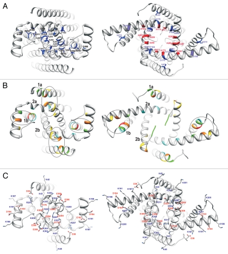
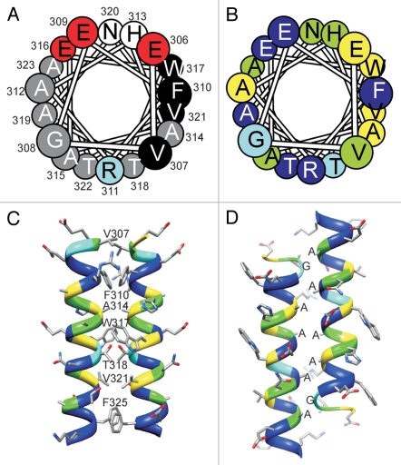
Mechanosensitive TREK channels belong to the family of K2P channels, a family of widely distributed, well modulated channels that uniquely have two similar or identical subunits, each with two TM1-P-TM2 motifs. Our goal is to build viable structural models of TREK channels, as representatives of K2P channels family. The structures available to be used as templates belong to the 2TM channels superfamily. These have low sequence similarity and different structural features: four symmetrically arranged subunits, each having one TM1-P-TM2 motif. Our model building strategy used two subunits of the template (KcsA) to build one subunit of the target (TREK-1). Our models of the Closed channel were adjusted to differ substantially from those of the template, e.g., TM2 of the 2nd repeat is near the axis of the pore whereas TM2 of the 1st repeat is far from the axis. Segments linking the two repeats and immediately following the last TM segment were modeled ab initio as α-helices based on helical periodicities of hydrophobic and hydrophilic residues, highly conserved and poorly conserved residues, and statistically related positions from multiple sequence alignments. The models were further refined by two-fold symmetry-constrained MD simulations using a protocol we developed previously. We also built models of the Open state and suggest a possible tension-activated gating mechanism characterized by helical motion with two-fold symmetry. Our models are consistent with deletion/truncation mutagenesis and thermodynamic analysis of gating described in the accompanying paper.


Figures

Figure 1
Figure 1
Modeling TREK-1 channel. (A) Schematic illustrating our approach to modeling TREK-1 channels: two KCSA subunits were used to model one TREK subunit; linker and C-terminal regions were modeled ab initio; helical segments are represented by cylinders. (B) Target-template sequence alignment used for modeling; identical residues are highlighted magenta, selectivity filter is highlighted orange; M1P1 loop was not included in the models; helical segments are underlined using the same color scheme as in (A). (C) Bottom view and (D) side view of structural models for the closed (left part), low-conductive (central part) and open (right part) conformation, in ribbon representation; helical segments are labeled and colored using the same color scheme as in (A and B).
Figure 2
Figure 2
Residue conservation patterns from multiple sequence alignments, used as a modeling criteria. (A) Target-template sequence alignment in which residues are highlighted according to their degree of conservation, on a scale from red (highly conserved) to blue (not conserved). (B) Top view and (C) bottom view of the models for closed (left), low-conductive (center) and open (right) conformations, colored according to the same degree-of-conservation scheme.
Figure 3
Figure 3
Ribbon representations of the inner pore region of closed (left) and open (right) models viewed from the inside. (A) Small residues located at positions of close contact between helices are colored blue. Small residues that become exposed in the lining of the pore when the channel opens are colored red. (B) High degree, high MI positions obtained from the MSA of TREK homologs, used as a modeling criteria. Residues with the highest degree values are colored according to the color spectrum scheme used in Supplementary Table 1, red for the highest score and cyan for the lowest. (C) Charged residues of the inner pore segments. Side chains are colored by element (gray = carbon, blue = nitrogen, red = oxygen, white = hydrogen). Positively charged resides are labeled in blue, negatively charged residues in red.
Figure 4
Figure 4
Models of the post-TM C-terminal segment (residues 305–330). (A) Helical wheel representation in which residues are colored according to polarity: small hydrophobic (grey); large hydrophobic (black); negatively charged (red) and positively charged (blue). (B) Helical wheel representation in which residues are colored according to their degree of conservation, from yellow (highly conserved) to blue (not conserved). (C) “Parallel Dimer” model in which the highly conserved hydrophobic residues self associate; this arrangement was used in the models of the open and closed states. (D) “Antiparallel Dimer” model in which the small residues face pack next to each other; this arrangement was used in a tentative model of the inactivated state.
Figure 5
Figure 5
Refinement of structural models by symmetry-restrained MD simulations. (A) Simulation protocol: models were subject to 30 ns unrestrained MD simulation (black line); after 5, 10, 15, and respectively 20 ns, a symmetry annealing step was applied (blue and green thick lines) followed by 10 ns unrestrained simulation (blue and green thin lines); the model annealed after 15 ns was subject to a second step of symmetry annealing followed by unrestrained simulations (red lines). (B) RMSD plots of the Cα trace of each model, during the first 10 ns of unrestrained simulation, using as reference the models at the beginning of each simulation step. (C) Root-mean square fluctuations (RMSF) of each model, during the last step of unrestrained simulation (thin red lines in A).

References

    1. Bayliss DA, Barrett PQ. Emerging roles for two-poredomain potassium channels and their potential therapeutic impact. Trends Pharmacol Sci. 2008;29:566–575. - PMC - PubMed
    1. Honoré E. The neuronal background K2P channels: focus on TREK-1. Nat Rev Neurosci. 2007;8:251–261. - PubMed
    1. Rajan S, Wischmeyer E, Karschin C, Preisig-Müller R, Grzeschik KH, Daut J, et al. THIK-1 and THIK-2, a novel subfamily of tandem pore domain K+ channels. J Biol Chem. 2001;276:7302–7311. - PubMed
    1. Lesage F, Lazdunski M. Molecular and functional properties of two-pore-domain potassium channels. Am J Physiol Renal Physiol. 2000;279:793–801. - PubMed
    1. Goldstein SA, Bockenhauer D, O'Kelly I, Zilberberg N. Potassium leak channels and the KCNK family of two-P-domain subunits. Nat Rev Neurosci. 2001;2:175–184. - PubMed

Publication types

MeSH terms

Substances

LinkOut - more resources