. 2011 Feb;131(2):401-9.
doi: 10.1038/jid.2010.319. Epub 2010 Nov 18.

Staphylococcus aureus hijacks a skin commensal to intensify its virulence: immunization targeting β-hemolysin and CAMP factor

Affiliations

Staphylococcus aureus hijacks a skin commensal to intensify its virulence: immunization targeting β-hemolysin and CAMP factor

Chih-Wei Lo et al. J Invest Dermatol. 2011 Feb.

Abstract

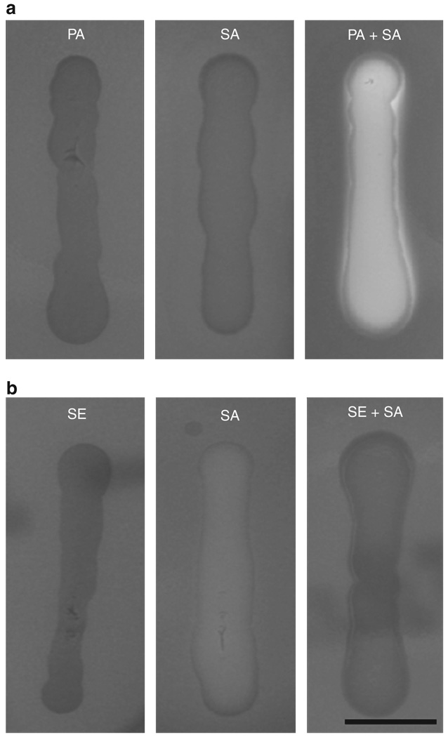
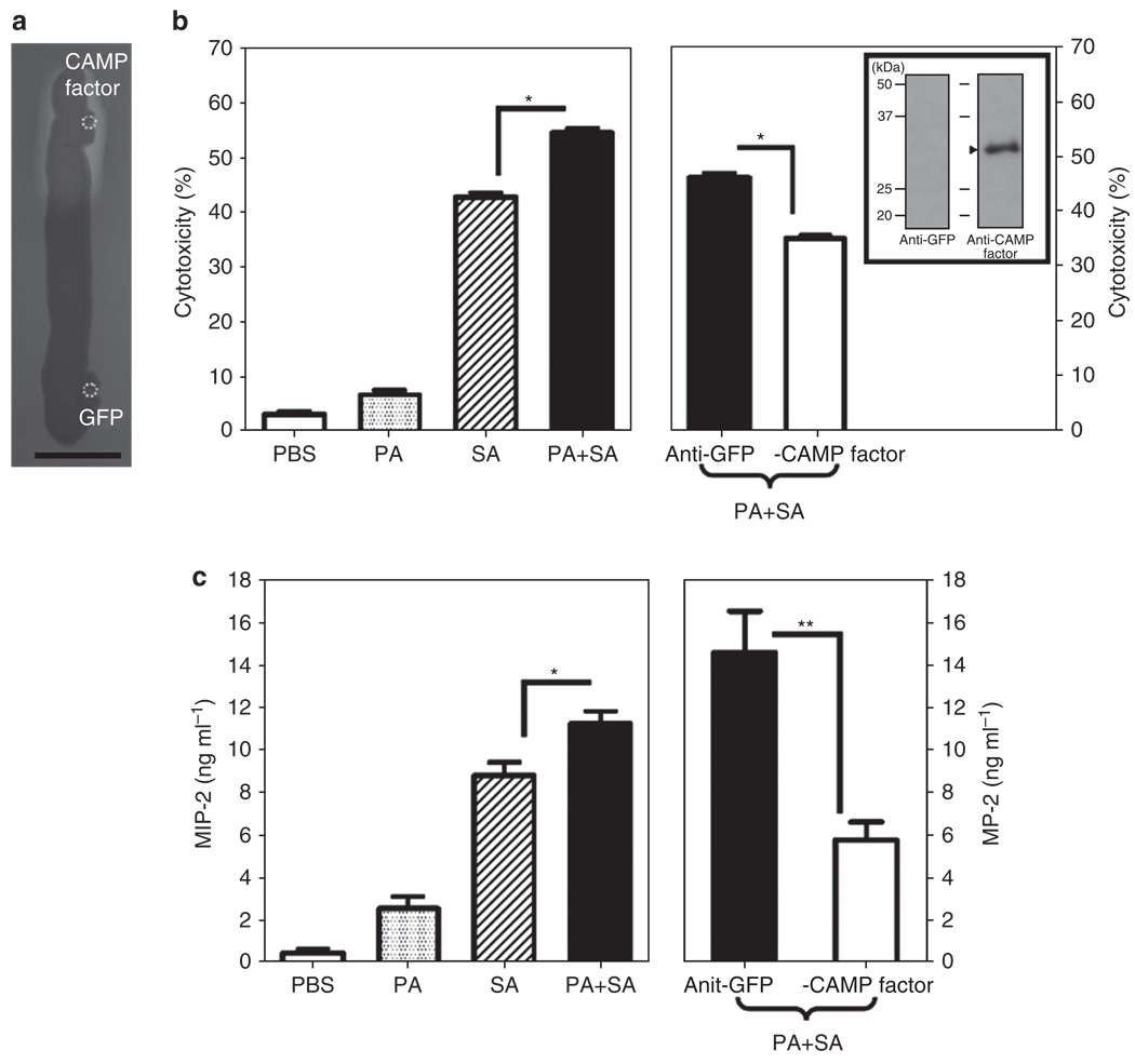
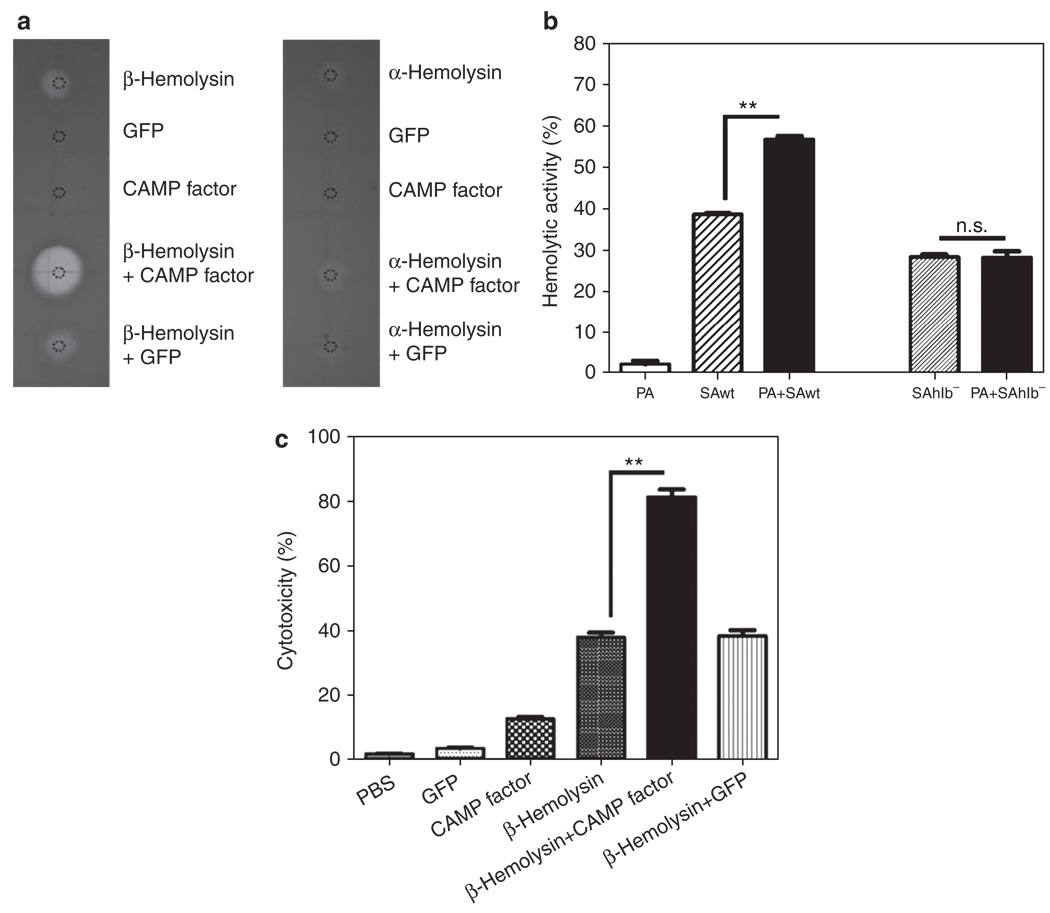
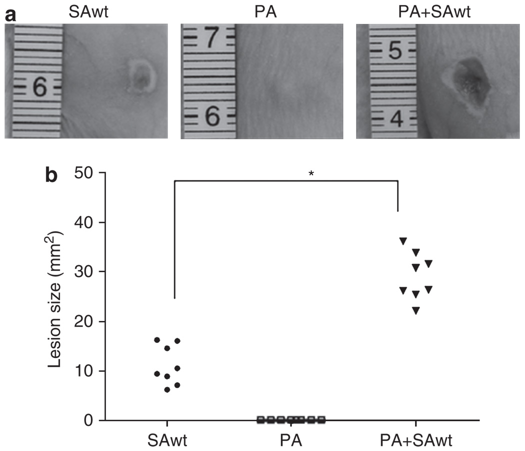
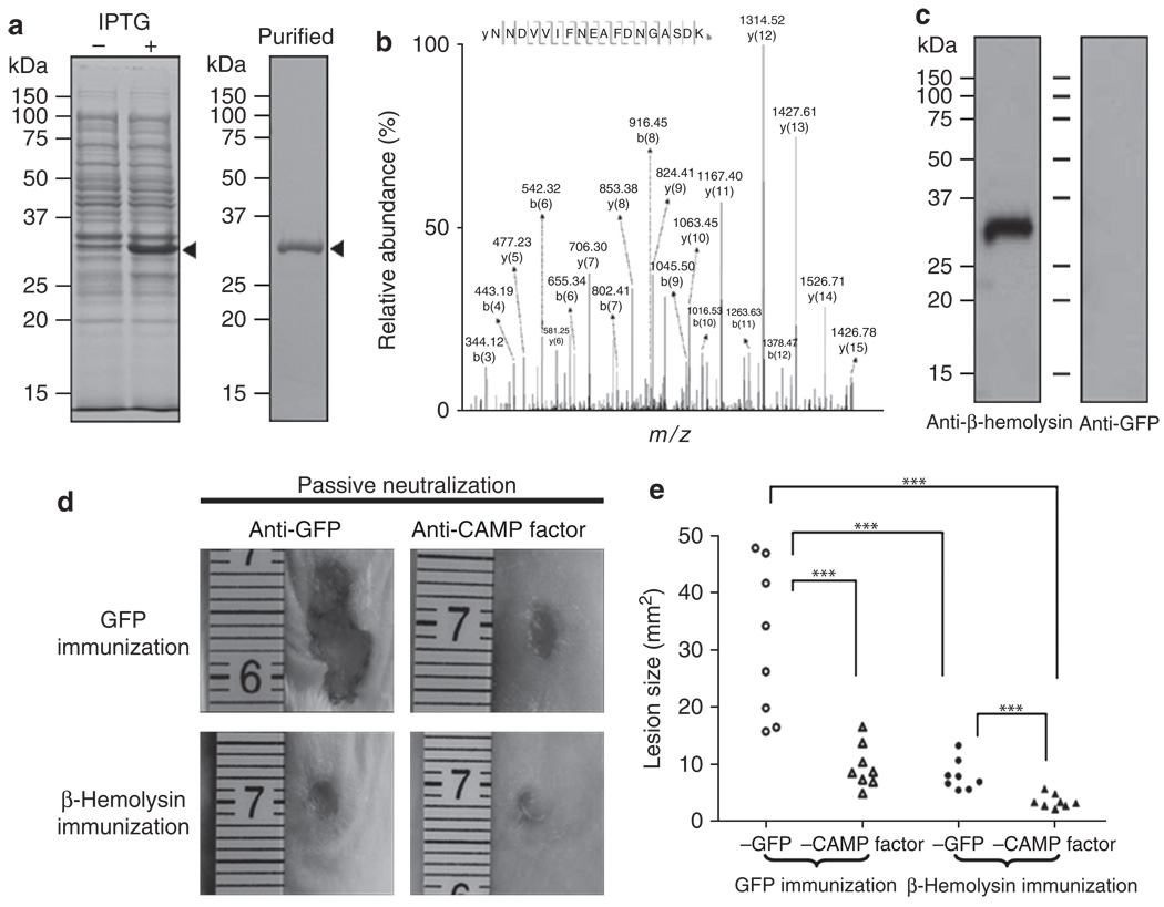
The need for a new anti-Staphylococcus aureus therapy that can effectively cripple bacterial infection, neutralize secretory virulence factors, and lower the risk of creating bacterial resistance is undisputed. Here, we propose what is, to our knowledge, a previously unreported infectious mechanism by which S. aureus may commandeer Propionibacterium acnes, a key member of the human skin microbiome, to spread its invasion and highlight two secretory virulence factors (S. aureus β-hemolysin and P. acnes CAMP (Christie, Atkins, Munch-Peterson) factor) as potential molecular targets for immunotherapy against S. aureus infection. Our data demonstrate that the hemolysis and cytolysis by S. aureus were noticeably augmented when S. aureus was grown with P. acnes. The augmentation was significantly abrogated when the P. acnes CAMP factor was neutralized or β-hemolysin of S. aureus was mutated. In addition, the hemolysis and cytolysis of recombinant β-hemolysin were markedly enhanced by recombinant CAMP factor. Furthermore, P. acnes exacerbated S. aureus-induced skin lesions in vivo. The combination of CAMP factor neutralization and β-hemolysin immunization cooperatively suppressed the skin lesions caused by coinfection of P. acnes and S. aureus. These observations suggest a previously unreported immunotherapy targeting the interaction of S. aureus with a skin commensal.


Conflict of interest statement

CONFLICT OF INTEREST

The authors state no conflict of interest.

Figures

Figure 1
Figure 1. Propionibacterium acnes, not Staphylococcus epidermidis, enhances the hemolytic activity of Staphylococcus aureus
(a) P. acnes alone (PA, 2 × 10⁷ colony-forming units (CFUs)), S. aureus 113 alone (SA, 2 × 10⁷ CFUs), P. acnes plus S. aureus (PA + SA, 1:1 ratio with a total of 2 × 10⁷ CFUs), (b) S. epidermidis alone (SE, 2 × 10⁷ CFUs), or S. epidermidis plus S. aureus (SE + SA, 1:1 ratio with a total of 2 × 10⁷ CFUs) were suspended in 10 µl PBS and streaked on sheep blood agar plates at 37°C for 2 days under anaerobic conditions. Hemolytic activity was determined as described in Materials and Methods. Bar = 1 cm. Data are representative of three separate experiments with similar results.
Figure 2
Figure 2. Propionibacterium acnes Christie, Atkins, Munch-Peterson (CAMP) factor mediates the enhancement of hemolysis and cytolysis caused by a coculture of P. acnes with Staphylococcus aureus
(a) A total of 10 µg of recombinant CAMP factor and green fluorescence protein (GFP) in 5 µl phosphate-buffered saline (PBS) was spotted (circles) adjacent to a S. aureus 113 (1 × 10⁵ colony-forming units (CFUs) in 10 µl PBS) streak on a sheep blood agar plate grown overnight at 37°C. Bar = 1 cm. (b) Macrophage RAW264.7 cells were treated with PBS or bacterial culture supernatants (10 µg ml−1) of P. acnes (PA), S. aureus (SA), or P. acnes plus S. aureus (PA + SA). Bacterial culture supernatants of coculture of P. acnes and S. aureus were incubated with 5% (v/v) anti-CAMP factor or anti-GFP (as a negative control) antiserum. The in vitro cytotoxicity assay was performed after cells were treated with the mixture of bacterial culture supernatant and antiserum for 24 hours. Western blot analysis was conducted to validate the specificity of anti-CAMP factor antiserum (arrowhead in the inset panel). One microgram of recombinant CAMP factor was separated via 12% SDS-PAGE, transferred to an Immobilon-P polyvinylidene difluoride membrane, and reacted with anti-CAMP factor (right lane) or anti-GFP antiserum (left lane), in 1:2,000 dilution. (c) Neutralization of CAMP factor significantly diminishes the enhancement of P. acnes on the S. aureus-induced macrophage inflammatory protein-2 (MIP-2) production. PBS (25 µl), P. acnes (PA, 25 µl; 2 × 10⁷ CFUs), S. aureus 113 (SA, 25 µl; 2 × 10⁷ CFUs), or a mixture of P. acnes plus S. aureus 113 (PA + SA, 25 µl; 1:1 ratio with a total of 2 × 10⁷ CFUs) was intradermally injected into the ears of imprinting control region (ICR) mice. The mixture of P. acnes and S. aureus (PA + SA) was preincubated with 5% (v/v) anti-CAMP factor or anti-GFP antiserum at 25°C for 1 hour to neutralize the P. acnes CAMP factor. After incubation, the bacteria alone or bacteria preincubated with antiserum were intradermally injected into ears of ICR mice. After injection for 24 hours, ears were excised, homogenized, and centrifuged. ELISA was performed to measure the proinflammatory MIP-2 cytokine in supernatant. The data are represented as mean ± SE (n = 3, *P<0.05 and **P<0.005 by Student’s t-test).
Figure 3
Figure 3. Staphylococcus aureus β-hemolysin contributes to the augmentation of hemolysis and cytolysis caused by coculture of Propionibacterium acnes and S. aureus
(a) Recombinant β-hemolysin (5 µg; Toxin Technology, Sarasota, FL), Christie, Atkins, Munch-Peterson (CAMP) factor (5 µg), and α-hemolysin (1 µg) in 5 µl phosphate-buffered saline (PBS) were spotted on sheep blood agar plates to examine the hemolytic activity. Recombinant green fluorescence protein (GFP; 5 µg) in 5 µl PBS was used as a control. (b) P. acnes (PA, 2 × 10⁷ colony-forming units (CFUs)) alone, wild-type S. aureus alone (SAwt, 2 × 10⁷ CFUs), or P. acnes plus wild-type S. aureus (1:1 ratio with a total of 2 × 10⁷ CFUs) was incubated with sheep blood cells at 37°C with end-over-end rotation for 2 days. Hemolytic activity was detected by measuring the absorbance of hemoglobin release at 540 nm. To examine the essentiality of S. aureus β-hemolysin in the enhancement of hemolysis by coculture of P. acnes and S. aureus, a β-hemolysin-deficient S. aureus (SAhlb, 2 × 10⁷ CFUs) was incubated with sheep blood cells (PA + SAhlb in 1:1 ratio with a total of 2 × 10⁷ CFUs) in the absence and presence of P. acnes. . (c) To determine the synergistic effect of CAMP factor and β-hemolysin on cytotoxicity, macrophage RAW264.7 cells (1 × 10⁵ per well) were incubated with 10 µl of 200 µg ml−1 recombinant proteins of GFP, CAMP factor, β-hemolysin, or the mixture of β-hemolysin plus CAMP factor or GFP at 37°C for 18 hours. An equal volume of PBS was used as a negative control. After incubation, the cytotoxicity of recombinant proteins to macrophages was determined by an in vitro cytotoxicity assay as described in Materials and Methods. Data are represented as mean ± SE (n = 3, **P<0.005 by Student’s t-test). NS, not significant.
Figure 4
Figure 4. Propionibacterium acnes intensifies Staphylococcus aureus-induced skin lesions
(a) Wild-type S. aureus (SAwt, 1 × 10⁷ colony-forming units (CFUs)), P. acnes (PA, 1 × 10⁷ CFUs), or P. acnes plus wild-type S. aureus (PA + SAwt, 1:1 ratio with a total of 2 × 10⁷ CFUs) in 50 µl phosphate-buffered saline was subcutaneously injected into the dorsal skins of imprinting control region mice to induce skin lesions. (b) Lesion sizes were examined and quantified 2 days after injection. Representative photographs of dorsal skin lesions are shown. Data are means of two independent experiments (n = 8, *P<0.05 by Student’s t-test).
Figure 5
Figure 5. The immunogenicity of β-hemolysin and the combination of β-hemolysin vaccination and Christie, Atkins, Munch-Peterson (CAMP) factor neutralization confer immune protection against bacteria-induced skin lesions
(a) Recombinant β-hemolysin (arrowheads) was expressed in E. coli BL21 (DE3). Competent cells transformed with the pEcoli-Nterm 6 × HN vector-inserted cDNA encoding β-hemolysin were incubated without (−) or with (+) isopropyl-β-d-thiogalactopyranoside (IPTG) for 4 hours and then subjected to 12% SDS-PAGE. Purified β-hemolysin is shown in the right panel. (b) The identity of recombinant β-hemolysin was analyzed by Nano liquid chromatography linear trap quadrupole tandem mass spectrometry (NanoLC-LTQ MS/MS). Tryptic digests of purified recombinant β-hemolysin were subjected to NanoLC-LTQ MS/MS. A sequenced internal peptide (NNDVVIFNEAFDNGASDK) of β-hemolysin is presented. The m/z value of each “y” and “b” ion in collision-induced dissociation spectra is indicated. All sequenced peptides are shown in Supplementary Table S1 online. (c) Immunogenicity of β-hemolysin was evaluated by western blotting. Imprinting control region mice were intranasally vaccinated with UV-inactivated E. coli BL21 (DE3) overexpressing β-hemolysin or green fluorescence protein (GFP). Anti-antisera were collected 1 week after second vaccination. One microgram of recombinant β-hemolysin was separated via 12% SDS-PAGE, transferred to an Immobilon-P polyvinylidene difluoride membrane, and reacted with anti-β-hemolysin (left lane) or anti-GFP antiserum (right lane), in 1:2,000 dilution. (d) The GFP-(upper row) and β-hemolysin-immunized mice (lower row) were used for evaluation of immune protection. Mice were injected subcutaneously with the mixture of wild-type Staphylococcus aureus and Propionibacterium acnes (1:1 ratio with a total of 2 × 10⁷ colony-forming units in 50 µl phosphate-buffered saline) that were preincubated with 5% (v/v) anti-CAMP factor antiserum (right panel) or anti-GFP antiserum (left panel) at 25°C for 1 hour. Lesion sizes were examined 2 days after injection. (e) Skin lesions were measured and statistics compiled. Representative photographs of dorsal skin lesions are shown. Data are from two independent replicated experiments (***P<0.001; Tukey’s honest significant difference comparing the four treatments, in a two-way analysis of variance on log-transformed data comparing the four treatments, with day included as a blocking factor).

Similar articles

Cited by

References

    1. Ahn CY, Ko CY, Wagar EA, et al. Microbial evaluation: 139 implants removed from symptomatic patients. Plast Reconstr Surg. 1996;98:1225–1229. - PubMed
    1. Bashir A, Mujahid TY, Jehan N. Antibiotic resistance profile: isolation and characterization of clinical isolates of staphylococci from patients with community-acquired skin infections. Pak J Pharm Sci. 2007;20:299–304. - PubMed
    1. Brook I, Frazier EH. Infections caused by Propionibacterium species. Rev Infect Dis. 1991;13:819–822. - PubMed
    1. Bubeck Wardenburg J, Schneewind O. Vaccine protection against Staphylococcus aureus pneumonia. J Exp Med. 2008;205:287–294. - PMC - PubMed
    1. Cogen AL, Nizet V, Gallo RL. Skin microbiota: a source of disease or defence? Br J Dermatol. 2008;158:442–455. - PMC - PubMed

Publication types

MeSH terms

LinkOut - more resources