

Refined human artificial chromosome vectors for gene therapy and animal transgenesis
- PMID: 21085194
- PMCID: PMC3125098
- DOI: 10.1038/gt.2010.147
Refined human artificial chromosome vectors for gene therapy and animal transgenesis
Abstract
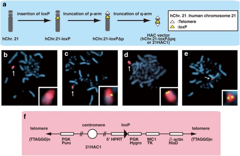
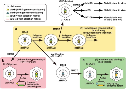
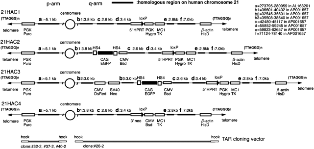
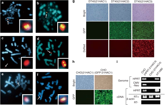
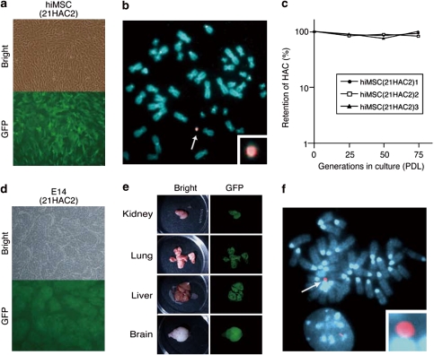
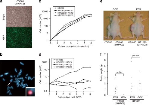
Human artificial chromosomes (HACs) have several advantages as gene therapy vectors, including stable episomal maintenance, and the ability to carry large gene inserts. We previously developed HAC vectors from the normal human chromosomes using a chromosome engineering technique. However, endogenous genes were remained in these HACs, limiting their therapeutic applications. In this study, we refined a HAC vector without endogenous genes from human chromosome 21 in homologous recombination-proficient chicken DT40 cells. The HAC was physically characterized using a transformation-associated recombination (TAR) cloning strategy followed by sequencing of TAR-bacterial artificial chromosome clones. No endogenous genes were remained in the HAC. We demonstrated that any desired gene can be cloned into the HAC using the Cre-loxP system in Chinese hamster ovary cells, or a homologous recombination system in DT40 cells. The HAC can be efficiently transferred to other type of cells including mouse ES cells via microcell-mediated chromosome transfer. The transferred HAC was stably maintained in vitro and in vivo. Furthermore, tumor cells containing a HAC carrying the suicide gene, herpes simplex virus thymidine kinase (HSV-TK), were selectively killed by ganciclovir in vitro and in vivo. Thus, this novel HAC vector may be useful not only for gene and cell therapy, but also for animal transgenesis.
Figures
Similar articles
-
Human artificial chromosome with a conditional centromere for gene delivery and gene expression.DNA Res. 2010 Oct;17(5):293-301. doi: 10.1093/dnares/dsq020. Epub 2010 Aug 26. DNA Res. 2010. PMID: 20798231 Free PMC article.
-
Human artificial chromosomes constructed using the bottom-up strategy are stably maintained in mitosis and efficiently transmissible to progeny mice.J Biol Chem. 2006 Sep 8;281(36):26615-23. doi: 10.1074/jbc.M603053200. Epub 2006 Jul 12. J Biol Chem. 2006. PMID: 16837455
-
A gene delivery system with a human artificial chromosome vector based on migration of mesenchymal stem cells towards human glioblastoma HTB14 cells.Neurol Res. 2010 May;32(4):429-37. doi: 10.1179/174313209X455718. Epub 2009 Jul 8. Neurol Res. 2010. PMID: 19589205
-
Developing de novo human artificial chromosomes in embryonic stem cells using HSV-1 amplicon technology.Chromosome Res. 2015 Feb;23(1):105-10. doi: 10.1007/s10577-014-9456-2. Chromosome Res. 2015. PMID: 25657030 Free PMC article. Review.
-
Transfer of human artificial chromosome vectors into stem cells.Reprod Biomed Online. 2008 Jan;16(1):57-69. doi: 10.1016/s1472-6483(10)60557-3. Reprod Biomed Online. 2008. PMID: 18252049 Review.
Cited by
-
Genetic therapy for the nervous system.Hum Mol Genet. 2011 Apr 15;20(R1):R28-41. doi: 10.1093/hmg/ddr110. Epub 2011 Mar 23. Hum Mol Genet. 2011. PMID: 21429918 Free PMC article. Review.
-
DNA-aptamer targeting vimentin for tumor therapy in vivo.Nucleic Acid Ther. 2014 Apr;24(2):160-70. doi: 10.1089/nat.2013.0471. Epub 2014 Jan 11. Nucleic Acid Ther. 2014. PMID: 24410722 Free PMC article.
-
Use of a Human Artificial Chromosome for Delivering Trophic Factors in a Rodent Model of Amyotrophic Lateral Sclerosis.Mol Ther Nucleic Acids. 2015 Oct 6;4(10):e253. doi: 10.1038/mtna.2015.28. Mol Ther Nucleic Acids. 2015. PMID: 26440597 Free PMC article.
-
Combinations of chromosome transfer and genome editing for the development of cell/animal models of human disease and humanized animal models.J Hum Genet. 2018 Feb;63(2):145-156. doi: 10.1038/s10038-017-0378-7. Epub 2017 Nov 27. J Hum Genet. 2018. PMID: 29180645 Review.
-
Highly Efficient Microcell-Mediated Transfer of HACs Containing a Genomic Region of Interest into Mammalian Cells.Curr Protoc. 2021 Sep;1(9):e236. doi: 10.1002/cpz1.236. Curr Protoc. 2021. PMID: 34491634 Free PMC article.
References
-
- O'Connor TP, Crystal RG. Genetic medicines: treatment strategies for hereditary disorders. Nat Rev Genet. 2006;7:261–276. - PubMed
-
- Hacein-Bey-Abina S, Von Kalle C, Schmidt M, McCormack MP, Wulffraat N, Leboulch P, et al. LMO2-associated clonal T cell proliferation in two patients after gene therapy for SCID-X1. Science. 2003;302:415–419. - PubMed
-
- Costantini F, Radice G, Lee JL, Chada KK, Perry W, Son HJ. Insertional mutations in transgenic mice. Prog Nucleic Acid Res Mol Biol. 1989;36:159–169. - PubMed
-
- Soriano P, Gridley T, Jaenisch R. Retroviruses and insertional mutagenesis in mice: proviral integration at the Mov 34 locus leads to early embryonic death. Genes Dev. 1987;1:366–375. - PubMed
Publication types
MeSH terms
LinkOut - more resources
Full Text Sources
Other Literature Sources
Medical
