. 2010 Nov 9;5(11):e13910.
doi: 10.1371/journal.pone.0013910.

Suppression of hypoxia-inducible factor 1α (HIF-1α) by tirapazamine is dependent on eIF2α phosphorylation rather than the mTORC1/4E-BP1 pathway

Affiliations

Suppression of hypoxia-inducible factor 1α (HIF-1α) by tirapazamine is dependent on eIF2α phosphorylation rather than the mTORC1/4E-BP1 pathway

Jun Zhang et al. PLoS One. .

Abstract

Hypoxia-inducible factor 1 (HIF-1), a heterodimeric transcription factor that mediates the adaptation of tumor cells and tissues to the hypoxic microenvironment, has attracted considerable interest as a potential therapeutic target. Tirapazamine (TPZ), a well-characterized bioreductive anticancer agent, is currently in Phase II and III clinical trials. A major aspect of the anticancer activity of TPZ is its identity as a tumor-specific topoisomerase IIα inhibitor. In the study, for the first time, we found that TPZ acts in a novel manner to inhibit HIF-1α accumulation driven by hypoxia or growth factors in human cancer cells and in HepG2 cell-derived tumors in athymic nude mice. We investigated the mechanism of TPZ on HIF-1α in HeLa human cervical cancer cells by western blot analysis, reverse transcription-PCR assay, luciferase reporter assay and small interfering RNA (siRNA) assay. Mechanistic studies demonstrated that neither HIF-1α mRNA levels nor HIF-1α protein degradation are affected by TPZ. However, TPZ was found to be involved in HIF-1α translational regulation. Further studies revealed that the inhibitory effect of TPZ on HIF-1α protein synthesis is dependent on the phosphorylation of translation initiation factor 2α (eIF2α) rather than the mTOR complex 1/eukaryotic initiation factor 4E-binding protein-1 (mTORC1/4E-BP1) pathway. Immunofluorescence analysis of tumor sections provide the in vivo evidences to support our hypothesis. Additionally, siRNA specifically targeting topoisomerase IIα did not reverse the ability of TPZ to inhibit HIF-1α expression, suggesting that the HIF-1α inhibitory activity of TPZ is independent of its topoisomerase IIα inhibition. In conclusion, our findings suggest that TPZ is a potent regulator of HIF-1α and provide new insight into the potential molecular mechanism whereby TPZ serves to reduce HIF-1α expression.


Conflict of interest statement

Competing Interests: The authors have declared that no competing interests exist.

Figures

Figure 1
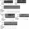
Figure 1. TPZ decreases hypoxia-induced HIF-1α protein accumulation.
HeLa cells (A), and HEK-293, OVCAR8, HepG2, SMMC-7721 and HCT116 cells (B) were exposed to hypoxia and a gradient of concentrations of TPZ for 4 h. HIF-1α and β-actin protein levels were detected by western-blot analysis of whole-cell extracts, as described in the Materials and Methods. (C) Effect of TPZ treatment on the expression level of HIF-1α in HepG2 cell-derived tumors, as determined by immunoblot analysis. (D) Densitometry analyses of (C). (E) Hypoxia-dependent HIF-1α transcriptional activity was measured using HRE-dependent reporter assays, as described in the Materials and Methods. HeLa cells were transiently transfected with the HRE-Luc plasmid and then treated with TPZ for 8 h under hypoxic conditions. Luminescence was measured and fold stimulation was obtained by normalizing the relative luciferase activity to that of untreated cells under hypoxic conditions.
Figure 2
Figure 2. TPZ does not affect HIF-1α mRNA expression or protein degradation, but decreases HIF-1α protein synthesis.
(A) HeLa cells were exposed to varying concentrations of TPZ for 4 h or a single concentration for the indicated times. Then, the total RNA was extracted and analyzed for HIF-1α mRNA expression by RT-PCR, using GAPDH as a control gene. (B) Cells exposed to hypoxia overnight were treated with cycloheximide (CHX) in the presence or absence of 20 µM TPZ for various periods, and HIF-1α protein levels were measured by western-blot analysis. HeLa cells were treated with TPZ, together with MG132 (C) or chloroquine diphosphate (CQ) (D), under the indicated conditions, followed by immunoblotting with anti-HIF-1α or anti-β-actin antibodies. Cells were pretreated for 30 min with MG132 and CQ to allow functional inhibition of the proteasome and lysosome. (E) HeLa cells were pre-incubated with CHX for 3 h in normoxic conditions and then placed in fresh medium and treated with or without 20 µM TPZ for the indicated times under hypoxic conditions. The cells were harvested and lysates were immunoblotted with an anti-HIF-1α antibody.
Figure 3
Figure 3. Effects of TPZ on Akt and the mTORC1 pathway in HeLa cells.
(A) Cells were cultured in hypoxia for 4 h in the presence of the indicated concentrations of TPZ before western-blotting analysis. (B) Immunoprecipitates prepared from the lysates of HeLa cells with Raptor antibody were used in kinase assays with full-length 4EBP1 as the substrate. (C) Immunoblotting was used to detect the phosphorylation of Akt at Ser473 or Thr308 after TPZ treatment. HeLa cells were transfected with siRNAs specifically targeting TSC2 (D) and 4E-BP1 (E) or control siRNAs, as described in the Materials and Methods. Transfected cells were incubated with or without 20 µM TPZ for 4 h under hypoxic conditions. Proteins were detected by western-blot analysis using specific antibodies.
Figure 4
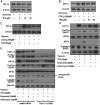
Figure 4. A direct role of eIF2α phosphorylation in HIF-1α downregulation by TPZ.
(A) HeLa cells treated with indicated concentrations of TPZ for 4 h under hypoxic conditions. Total lysates were probed for expression of HIF-1α, p-eIF2α, and eIF2α, while β-actin served as a loading control. (B) HeLa cells were exposed to varying concentrations of TPZ for 4 h. GADD153 mRNA levels were determined by real-time PCR. The relative fold change of GADD153 mRNA compared to GAPDH mRNA in untreated cells under hypoxia was arbitrarily set as 1.0. (C) The effect of TPZ treatment to basal protein synthesis was measured using ODD-dependent reporter assays. HeLa cells were transiently transfected with the ODD-Luc plasmid and then treated with TPZ for 4 h under normoxia or hypoxia. Luminescence was measured and fold stimulation was obtained by normalizing the relative luciferase activity of cells cultured under hypoxic conditions to those of untreated cells cultured under normoxic conditions. (D) Cells were untransfected or transfected with eIF2α-targeting siRNA for two days, followed by treatment with or without 20 µM TPZ. Western blotting of cell lysates was performed using the indicated antibodies.
Figure 5
Figure 5. TPZ reduces HIF-1α protein only under hypoxic conditions, and its activity is independent of its topoisomerase II inhibition.
(A) HeLa cells were exposed to normoxic conditions for 4 h in the presence of TPZ. (B) Cells were treated with varying concentrations of TPZ for 4 h, as indicated, in the presence of the hypoxia-mimetic agent cobalt chloride (CoCl2). (C) HeLa cells were treated for 4 h in normoxia, hypoxia or with CoCl2 in the presence of TPZ at the indicated concentrations. (D) Cells were treated with 20 µM TPZ, 20 µM etoposide and 10 µM adriamycin for 4 h. (E) Topoisomerase IIα was knocked down using siRNA, as described in the Materials and Methods, followed by drug treatment (20 µM TPZ, 20 µM etoposide and 10 µM adriamycin). Proteins were detected by western-blot analysis.
Figure 6
Figure 6. The effect of TPZ on HepG2 human xenograft models.
The mice transplanted with HepG2 human xenograft were randomly divided into two groups and given injection of TPZ (30 mg/kg) or vehicle every 2 days. Representative photomicrographs (magnification, x100) showing immunofluorescence staining for HIF-1α (A) and p-eIF2α (B) in tumor sections of vehicle–treated and TPZ-treated mice. The immunostaining data were confirmed in two or more specimens of each group. (C) Tumor volume are expressed as mean ± SD. (D) Relative tumor volume are expressed as mean ± SD. (**p<0.01, relative to vehicle group).

Similar articles

Cited by

References

    1. Li SH, Shin DH, Chun YS, Lee MK, Kim MS, et al. A novel mode of action of YC-1 in HIF inhibition: stimulation of FIH-dependent p300 dissociation from HIF-1{alpha}. Mol Cancer Ther. 2008;7:3729–3738. - PubMed
    1. Wouters BG, Koritzinsky M. Hypoxia signalling through mTOR and the unfolded protein response in cancer. Nat Rev Cancer. 2008;8:851–864. - PubMed
    1. Zhang H, Qian DZ, Tan YS, Lee K, Gao P, et al. Digoxin and other cardiac glycosides inhibit HIF-1alpha synthesis and block tumor growth. Proc Natl Acad Sci U S A. 2008;105:19579–19586. - PMC - PubMed
    1. Lou JJ, Chua YL, Chew EH, Gao J, Bushell M, et al. Inhibition of hypoxia-inducible factor-1alpha (HIF-1alpha) protein synthesis by DNA damage inducing agents. PLoS One. 2010;5:e10522. - PMC - PubMed
    1. Lau CK, Yang ZF, Lam CT, Tam KH, Poon RT, et al. Suppression of hypoxia inducible factor-1alpha (HIF-1alpha) by YC-1 is dependent on murine double minute 2 (Mdm2). Biochem Biophys Res Commun. 2006;348:1443–1448. - PubMed

Publication types

MeSH terms