. 2010 Nov 9;5(11):e13890.
doi: 10.1371/journal.pone.0013890.

Early acquisition of neural crest competence during hESCs neuralization

Affiliations

Early acquisition of neural crest competence during hESCs neuralization

Carol Lynn Curchoe et al. PLoS One. .

Abstract

Background: Neural crest stem cells (NCSCs) are a transient multipotent embryonic cell population that represents a defining characteristic of vertebrates. The neural crest (NC) gives rise to many derivatives including the neurons and glia of the sensory and autonomic ganglia of the peripheral nervous system, enteric neurons and glia, melanocytes, and the cartilaginous, bony and connective tissue of the craniofacial skeleton, cephalic neuroendocrine organs, and some heart vessels.

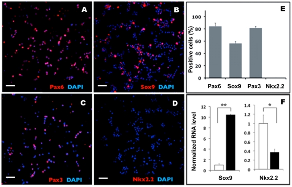
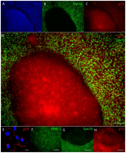
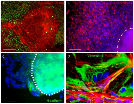
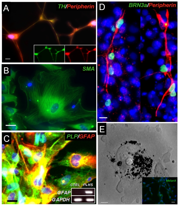
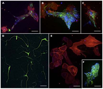
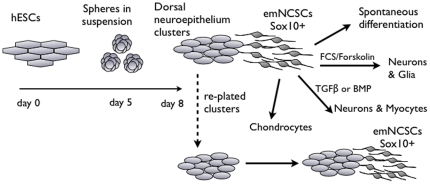
Methodology/principal findings: We present evidence that neural crest (NC) competence can be acquired very early when human embryonic stem cells (hESCs) are selectively neuralized towards dorsal neuroepithelium in the absence of feeder cells in fully defined conditions. When hESC-derived neurospheres are plated on fibronectin, some cells emigrate onto the substrate. These early migratory Neural Crest Stem Cells (emNCSCs) uniformly upregulate Sox10 and vimentin, downregulate N-cadherin, and remodel F-actin, consistent with a transition from neuroepithelium to a mesenchymal NC cell. Over 13% of emNCSCs upregulate CD73, a marker of mesenchymal lineage characteristic of cephalic NC and connexin 43, found on early migratory NC cells. We demonstrated that emNCSCs give rise in vitro to all NC lineages, are multipotent on clonal level, and appropriately respond to developmental factors. We suggest that human emNCSC resemble cephalic NC described in model organisms. Ex vivo emNCSCs can differentiate into neurons in Ret.k(-) mouse embryonic gut tissue cultures and transplanted emNCSCs incorporate into NC-derived structures but not CNS tissues in chick embryos.

Conclusions/significance: These findings will provide a framework for further studying early human NC development including the epithelial to mesenchymal transition during NC delamination.


Conflict of interest statement

Competing Interests: The authors have declared that no competing interests exist.

Figures

Figure 1
Figure 1. Neuralized human ES cell-derived express markers of dorsal neuroepithelium.
hESCs were neuralized for 5 days in floating spheres, dissociated and cytospins were immunostained for Pax6 (A), Sox9 (B), Pax3 (C), and Nkx2.2 (D). (E) Quantification of the results in A–D. (F) Q-PCR analysis of Sox9 and NKX2.2 expression in day 5 spheres (black bars) vs hESC (white bars). All values normalized to 18S expression; * = p<0.05, ** = p<0.0001, one-tail unpaired t-test. Scale bars 50 µM.
Figure 2
Figure 2. Early migratory NCSCs upregulate Sox10.
Immunostaining of adherent neurospheres and emigrating NCSCs after 3 days on fibronectin-coated plates. (A) DAPI staining for nuclei. Note the rosette structures within the adherent neurospheres. (B) Sox10 staining is observed in emigrating cells, but not in the adherent neurospheres. (C) p75 staining is observed in adherent neurospheres and emigrating cells. (D) High magnification of the overlay in B–C. (E) Confocal image of emigrating NCSCs confirms the membrane localization of p75. (F) HNK-1 staining is observed in adherent neurospheres and emigrating cells. (G, H) Adherent neurospheres were manually removed and replated on fibronectin and the second wave of emigrating cells was observed. (G) Secondary emigrating cells acquire Sox10, while the majority of cells in the replated neurosphere are Sox10-negative. (H) The expression of in the second wave of emigrating cells. Scale bars in A, B, C and D represent 100 µm and 10 µm in E Bars in F,G and H depict 150 µm.
Figure 3
Figure 3. Epithelial to mesenchymal transition in migratory emNCSCs.
(A) Phalloidin stains thick bands of f-actin within the adherent Sox10-negative neurosphere and thin cell border bands. Diffuse f-actin staining is visible in migratory Sox10-positive cells. (B) Connexin 43 (red) is acquired in migratory emNCSCs. Attached sphere is outlined. (C) N-Cadherin (green) is present in adherent neurospheres and gradually downregulated in migratory emNCSCs visualized by the nuclear DAPI IHC. (D) Confocal image of thick f-actin (red) bands at the border of an adherent neurosphere (outlined). Vimentin (green) is acquired in emNCSCs migrating away from the attached neurosphere. Scal bars in A and B represent 50 µm, in C 25 µm, and in D 10 µm. Cell nuclei were visualized by DAPI unless indicated differently.
Figure 4
Figure 4. Microarray analysis of the adherent neurospheres and emNCSCs.
The top most upregulated gene transcripts in emNCSCs (red) and neuroepithelial clusters (blue) are shown. The middle insert highlights the selected set of NC-related genes upregulated in emNCSCs.
Figure 5
Figure 5. p75 expression doesn't discriminate emNCSCs from neuroepithelial clusters.
(A) Day 8 cultures (containing emNCSC and neuroepithelial clusters) can be FACS-sorted based on p75 expression defining three populations: negative, medium and highly positive for p75. The grey profile corresponds to control situation with the secondary antibody only. (B) All fractions of cells were positive for Sox10 (85, 70 and 60% respectively for high, medium and negative). (C) All fractions can differentiate into neuronal, glial and smooth muscle lineages (p75-negative fraction is shown as an example). p75-negative sorted cells are reactive for; I. GFAP, II. β-3-tubulin, III. nestin, IV. smooth muscle actin. Cell nuclei are visualized by DAPI. Magnifications are 100x. Scale bars 30 µM.
Figure 6
Figure 6. Spontaneous differentiation of emNCSCs.
(A) Peripheral neuron-like cells are co-stained with TH (green) and Peripherin (red). Individual channels are shown in the inserts.(B) Smooth muscle cells reactive to smooth muscle actin. (C) The addition of 1% horse serum to the NS medium yielded GFAP-positive cells co-expressing p75NTR and proteolipid protein (PLP), consistent with Schwann cell identity. Inset, PCR detection of GFAP transcript. CTRL  =  NS medium. Scale bar  = 25 ìm. (D) Brn3a (red) and Peripherin (green) staining indicates differentiation into peripheral sensory-like neurons. (E) Bright field image of black inclusion bodies in a characteristic melanosome, inset box shows Melan A (green) reactivity. Nuclei were visualized by DAPI. Scale bars represent 40 µm in A, C and D, 60 µm in D, and 100 µm in E and its inset.
Figure 7
Figure 7. Directed differentiation of emNCSCs.
The growth factors FGF, EGF, insulin, and nicotinamide were withdrawn from NS medium and the base medium is supplemented with TGFβ (A-D) to enrich differentiating cells for neuronal fates, FCS/Forskolin to enrich for neuro-glial fates (E–H) and BMP2 to enrich for smooth muscle (I–L). A, E and I show GFAP immunohistochemistry (green, astrocytes). B, F and J indicate bIII tubulin (green, immature neurons). C, G and K show nestin (red, neuroblasts) and D, H and L show SMA reactivity (green, smooth muscle actin). Cell nuclei were visualized by DAPI, bars in A–L represent 100 µm. (M,N) Chondrocytic differentiation can be directed in emNCSC cultures (see materials and methods for details) and cryosectioned pellets reveal collagen II by Alcian blue staining. Scale bars 100 µM in M and 25 µM in N.
Figure 8
Figure 8. Clonal analysis of emNCSC.
Representative examples of multipotent (A–C), bi-potent (F), and unipotent (D, E) clones of emNCSC. For all images: TuJ1 (green), SMA (red), GFAP (blue); Scale bars 50 µM.
Figure 9
Figure 9. NC competency is ablated by treatment with Noggin or DKK.
The effect of Noggin (BMP inhibitor) and DKK (Wnt inhibitor) on hESC-derived emNCSCs. Untreated (control) neurosphere cultures are strongly reactive for (A) SOX10 (green), (B) HNK-1 (green), and (C) p75 (red) and negative for the CNS marker (D) Olig2 (red). The addition of Noggin to NS medium ablates (E) SOX10 (green), (F) HNK-1 (green), and (G) p75 (red) and induces expression of the CNS marker (H) Olig2 (red). Culturing neurospheres from day 0 in media that was collected from 293T cells transfected with a DKK plasmid ablates expression of the NC markers (I) SOX10 and (J) HNK-1 while the low affinity neutrophin receptor (K) p75 was still expressed. The insets in upper left corners show the cell nuclei (DAPI). Scale bars represent 50 µm in A, and C–H, 10 µm in B, and 100 µm in I–K.
Figure 10
Figure 10. CD73 expression in emNCSCs.
Early migratory NC cells are considered to be a uniform population of cells capable of differentiation into mesenchymal and PNS derivatives. To evaluate the potential of hESC-derived emNCSCs the mesenchymal marker CD73 was investigated at several time points, followed by prospective isolation and differentiation. (A) Day 5 neurospheres were tested for CD73 expression, approximately 0.11% of cells were positive. (B) Adherent neuroepithelial clusters were mechanically separated from migrating cells and tested for CD73 expression, approximately 0.5% of adherent neuroepithelial rosettes cells were positive. (C) 13% of cells migrating from neuroepithelial rosettes (SOX10-positive cells) are positive for CD73. (D) Migrating cells were sorted based on CD73 expression and cultured for 1 week. Both CD73-positive and -negative cells yielded nestin- and SMA-positive cells (areas with abundant staining are shown), however, the MAP2 reactive cells were enriched 10-fold in the CD73-negative fraction. Both populations had rare GFAP-positive cells and scattered KI67-positive cells. Scale bars represent 10 µm in I, I′ and V′, 50 µm in III, IV, III′ and IV′, and 100 µm in II, V and II′.
Figure 11
Figure 11. Injection of emNCSCs in aganglionic bowels in ex vivo organotypic cultures.
EmNCSCs can colonize aganglionic guts in organotypic gut cultures and differentiate into neurons. (A) Confocal image of a cryosectioned Ret KO gut segment 7 days post injection labeled with fluorescent antibodies against neurofilament (red). (B) Non-injected Ret KO gut segment 7 days post injection showing no neurofilament (red) reactivity. (C) In heterozygous animals endogenous neurons are present in the non-injected gut cultures (neurofilament staining in red). Cell nuclei are visualized by DAPI. Scale bars in A-C,10 µm. (D) Whole mount of a Ret KO gut segment stained with a neurofilament marker (red) and anti-GFP expressed by emNCSC (green). The GFP staining overlaps with the neurofilament staining in several places showing emNCSC-derived neurons; one example is marked by asterisk. Single channels (red, neurofilament and green, GFP) are depicted on top of the merged photograph for better identification (Magnification 20x).
Figure 12
Figure 12. Injection of emNCSCs into the cranial NC pathway of chick embryos.
(A) Whole mount view of a clump of grafted emNCSCs (arrow) injected into the cranial region of a stage 11 chick embryo. (B–H) Sections through the cranial region of chick embryos 5 days post-injection labeled with fluorescent antibodies to human nuclei (HuN, green), HNK-1 (blue), neuronal marker HuC/HuD (purple) and smooth muscle actin (SMA, red). (B) A number of human emNCSCs remain clustered and express HNK-1 (arrows) while individual cells that have left the graft site (arrowheads) are negative for HNK-1 and contribute to head mesenchyme. (C) emNCSCs along HNK-1-positive nerves (arrowheads). (D) SMA-positive (arrow) and -negative (arrowhead) emNCSCs around a blood vessel. (E) HNK-1-positive emNCSCs within skeletal muscle (arrowhead) do not express muscle actin (red). (F) Some emNCSCs (arrows, left and right panels) in the TG express the neuronal marker HuC/HuD (middle and right panels). (G) emNCSCs in undifferentiated mesenchyme near blood vessels around the perimeter of the midbrain. No emNCSCs were observed in the midbrain. (H) Differentiated emNCSCs (left and right panels) expressing SMA (arrow, middle and right panels) or HNK-1 (arrowhead, middle and right panels). BV - blood vessel; TG – trigeminal ganglion. Scale bars 200 µM in A, 100 µM in B, 20 µM in C–H.
Figure 13
Figure 13. Scheme of early emNCSCs derivation from hESCs and differentiation into NC lineages.
hESC clusters are suspended for 5 days in NS medium. On day 5 of suspension culture neurospheres were allowed to adhere on fibronectin-coated plates and cultured in NS medium for 3 more days. Adherent neurospheres composed of neuroepithelial rosettes are negative for Sox10 and CD73. Migrating cells are positive for the NC markers Sox10 and CD73 (13%). Both cell types are positive for p75 and HNK-1. Rosettes can be manually excised and replated (dotted arrow) to yield additional waves of Sox10-positive emNCSCs. The adherent rosettes are therefore competent to become NC after they have undergone the EMT. Migratory emNCSCs can be allowed to spontaneously acquire NC lineage fates or can be directed to enrich for certain cell fates by the addition of growth factors.

Similar articles

Cited by

References

    1. Morrison SJ, White PM, Zock C, Anderson DJ. Prospective identification, isolation by flow cytometry, and in vivo self-renewal of multipotent mammalian neural crest stem cells. Cell. 1999;96:737–749. - PubMed
    1. Wong CE, Paratore C, Dours-Zimmermann MT, Rochat A, Pietri T, et al. Neural crest-derived cells with stem cell features can be traced back to multiple lineages in the adult skin. J Cell Biol. 2006;175:1005–1015. - PMC - PubMed
    1. Yoshida S, Shimmura S, Nagoshi N, Fukuda K, Matsuzaki Y, et al. Isolation of multipotent neural crest-derived stem cells from the adult mouse cornea. Stem Cells. 2006;24:2714–2722. - PubMed
    1. Kawaguchi J, Nichols J, Gierl MS, Faial T, Smith A. Isolation and propagation of enteric neural crest progenitor cells from mouse embryonic stem cells and embryos. Development. 2010;137:693–704. - PMC - PubMed
    1. LaBonne C, Bronner-Fraser M. Molecular mechanisms of neural crest formation. Annu Rev Cell Dev Biol. 1999;15:81–112. - PubMed

Publication types

MeSH terms

Substances