. 2010 Nov 15;5(11):e15507.
doi: 10.1371/journal.pone.0015507.

Role of the epigenetic regulator HP1γ in the control of embryonic stem cell properties

Affiliations

Role of the epigenetic regulator HP1γ in the control of embryonic stem cell properties

Maïa Caillier et al. PLoS One. .

Abstract

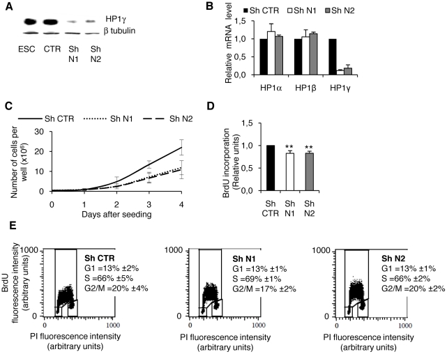
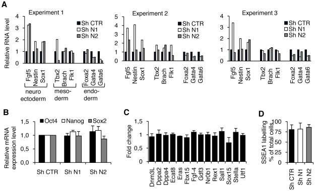
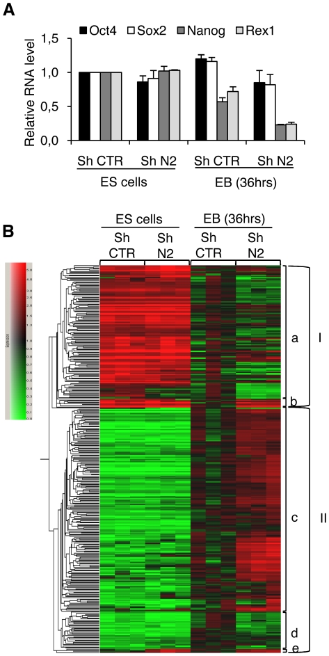
The unique properties of embryonic stem cells (ESC) rely on long-lasting self-renewal and their ability to switch in all adult cell type programs. Recent advances have shown that regulations at the chromatin level sustain both ESC properties along with transcription factors. We have focused our interest on the epigenetic modulator HP1γ (Heterochromatin Protein 1, isoform γ) that binds histones H3 methylated at lysine 9 (meH3K9) and is highly plastic in its distribution and association with the transcriptional regulation of specific genes during cell fate transitions. These characteristics of HP1γ make it a good candidate to sustain the ESC flexibility required for rapid program changes during differentiation. Using RNA interference, we describe the functional role of HP1γ in mouse ESC. The analysis of HP1γ deprived cells in proliferative and in various differentiating conditions was performed combining functional assays with molecular approaches (RT-qPCR, microarray). We show that HP1γ deprivation slows down the cell cycle of ESC and decreases their resistance to differentiating conditions, rendering the cells poised to differentiate. In addition, HP1γ depletion hampers the differentiation to the endoderm as compared with the differentiation to the neurectoderm or the mesoderm. Altogether, our results reveal the role of HP1γ in ESC self-renewal and in the balance between the pluripotent and the differentiation programs.


Conflict of interest statement

Competing Interests: The authors have declared that no competing interests exist.

Figures

Figure 1
Figure 1. HP1γ knockdown ESC exhibit an altered proliferation rate.
A. Two different shRNA directed against HP1γ (shN1 and shN2) were compared to a control shRNA (shCTR). Knockdown efficiency was measured by western blot using an anti-HP1γ antibody and compared to the basal level expressed in untransduced ESC and in shCTR cells. Beta-tubulin was used as a protein loading control. B. mRNA levels of the three HP1 isoforms HP1α, β and γ were measured by RT-qPCR. Values are represented relative to the ones obtained from shCTR-transduced cells. C. Growth curves for the shCTR, shN1 and shN2 cell lines representing the number of cells obtained after 1, 2, 3 or 4 days of culture. D. Proliferation measured by BrdU incorporation. BrdU was added to exponentially proliferating cells during the last 8 hours of culture. Results are represented relative to the value obtained in the shCTR cell line. Results are the means of three independent experiments. ** p<0,002 by t test. E. Cell cycle analysis using propidium iodide (PI) and BrdU incorporation. All values are means +/− SD from three independent experiments.
Figure 2
Figure 2. HP1γ knocked-down ESC sporadically express differentiation markers but show normal expression of pluripotency markers.
A. The expression of the indicated genes representing the three germ layers (neuroectoderm, mesoderm and endoderm) was measured by RT-qPCR. Results from three independent RNA samples are reported to illustrate the variability of the results obtained when genes are analyzed individually. B. The RNA expression levels of the core set of transcription factors required to sustain pluripotency was measured in shRNA cell lines by real time PCR. C. Fold change of additional markers associated with pluripotency obtained from microarrays analysis. Results are mean +/− SD of three independent microarrays experiments. D. The proportion of undifferentiated cells in the three shRNA cell lines was measured by immunolabelling of SSEA1 and subsequent flow cytometry analysis. In A, B and C results are reported as a ratio of the values obtained in the shCTR cells. For B and D, the means and standard errors were calculated from four independent experiments.
Figure 3
Figure 3. HP1γ knockdown increases the proportion of cells expressing the differentiation marker Brachyury.
ESC transfected with a Brachyury promoter-GFP construct were further transfected with the three shRNA. The promoter activity was followed by GFP expression measured by flow cytometry. A. The proportion of GFP positive cells is reported as the ratio of the value obtained in the shCTR cells. B. Representation of the mean fluorescence intensities (MFI) in GFP positive cells. The means and standard errors were calculated from four independent experiments. *** p<0.02 by t test.
Figure 4
Figure 4. A low level of HP1γ favors ESC commitment to differentiation.
The three shRNA cell lines (shN1, shN2 and shCTR) were seeded at low density and cultured with medium containing the indicated concentrations of LIF; the concentration of 1000 U/ml corresponds to that used in proliferating medium. Five days after seeding, colonies were fixed, stained for alkaline phosphatase and scored as undifferentiated (AP+), mixed (AP+/−) or differentiated (AP-). For each concentration of LIF, results are represented as the percent of the total number of colonies. Means and standard errors were calculated from three independent experiments, and t tests were performed to determine the significance of the differences between shHP1 and shCTR cells at each LIF concentration for a given type of colony (AP+, AP- or mixed). * p<0.1, ** p<0.05, *** p<0.02.
Figure 5
Figure 5. HP1γ knockdown improves early differentiation of ESC.
The transcriptome of shN2 cells was compared to that of shCTR cells using Affymetrix chip hybridization. RNA were isolated from cells that were cultivated in the absence of LIF and in non-adherent conditions to form embryoid bodies (EB) over the course of 36 hours. A. RNA levels of the three pluripotent transcription factors measured by real time PCR in the same RNA extracts that were compared by microarrays analysis. B. Variation of genes associated with differentiation are represented relative to the mean value obtained in undifferentiated shCTR RNA. Upregulated expression is indicated in red, downregulated expression is indicated in green; the variations obtained from the three independent differentiation experiments are represented.
Figure 6
Figure 6. HP1γ deprivation of ESC specifies differentiation during embryoid bodies formation.
The three shRNA cell lines (shN1, shN2 and shCTR) were induced to differentiate by embryoid bodies formation. Differentiation efficiency was assessed following the decrease of pluripotency markers (purple). Transcript levels of markers representing the three germ layers (neuroectoderm in blue, mesoderm in red and endoderm in green) were measured by RT-qPCR each day (d) after seeding. Results represented relatively to the value obtained in ESC (d0), are the mean of duplicates +/− SD from one experiment representative of two.
Figure 7
Figure 7. Specification of ESC differentiation by HP1γ deprivation is also observed in retinoic acid-induced cells.
The three shRNA cell lines (shN1, shN2 and shCTR) were induced to differentiate using retinoic acid. Differentiation efficiency was assessed following the decrease of pluripotency markers (in purple). Transcript levels of markers representing the three germ layers (neuroectoderm in blue, mesoderm in red and endoderm in green) were measured by RT-qPCR after 2 or 6 days of differentiation. Results represented relatively to the value obtained in ESC (d0), are the mean of duplicates +/− SD from one experiment representative of three.
Figure 8
Figure 8. HP1γ protein level remains constant during ESC differentiation.
ESC cells were induced to differentiate using 100 nM retinoic acid for 1 to 8 days (upper figure) or by embryoid bodies formation for 1 to 5 days (lower figure). The expression of the HP1γ, Oct4 and Nanog proteins were observed by western blot. Beta-tubulin was used as a protein loading control.

Similar articles

Cited by

References

    1. O'Shea KS. Self-renewal vs. differentiation of mouse embryonic stem cells. Biol Reprod. 2004;71:1755–1765. - PubMed
    1. Nichols J, Zevnik B, Anastassiadis K, Niwa H, Klewe-Nebenius D, et al. Formation of pluripotent stem cells in the mammalian embryo depends on the POU transcription factor Oct4. Cell. 1998;95:379–391. - PubMed
    1. Niwa H, Miyazaki J, Smith AG. Quantitative expression of Oct-3/4 defines differentiation, dedifferentiation or self-renewal of ES cells. Nat Genet. 2000;24:372–376. - PubMed
    1. Chambers I, Colby D, Robertson M, Nichols J, Lee S, et al. Functional expression cloning of Nanog, a pluripotency sustaining factor in embryonic stem cells. Cell. 2003;113:643–655. - PubMed
    1. Mitsui K, Tokuzawa Y, Itoh H, Segawa K, Murakami M, et al. The homeoprotein Nanog is required for maintenance of pluripotency in mouse epiblast and ES cells. Cell. 2003;113:631–642. - PubMed

Publication types

Substances