. 2010 Nov 15;5(11):e13989.
doi: 10.1371/journal.pone.0013989.

APP processing induced by herpes simplex virus type 1 (HSV-1) yields several APP fragments in human and rat neuronal cells

Affiliations

APP processing induced by herpes simplex virus type 1 (HSV-1) yields several APP fragments in human and rat neuronal cells

Giovanna De Chiara et al. PLoS One. .

Abstract

Lifelong latent infections of the trigeminal ganglion by the neurotropic herpes simplex virus type 1 (HSV-1) are characterized by periodic reactivation. During these episodes, newly produced virions may also reach the central nervous system (CNS), causing productive but generally asymptomatic infections. Epidemiological and experimental findings suggest that HSV-1 might contribute to the pathogenesis of Alzheimer's disease (AD). This multifactorial neurodegenerative disorder is related to an overproduction of amyloid beta (Aβ) and other neurotoxic peptides, which occurs during amyloidogenic endoproteolytic processing of the transmembrane amyloid precursor protein (APP). The aim of our study was to identify the effects of productive HSV-1 infection on APP processing in neuronal cells. We found that infection of SH-SY5Y human neuroblastoma cells and rat cortical neurons is followed by multiple cleavages of APP, which result in the intra- and/or extra-cellular accumulation of various neurotoxic species. These include: i) APP fragments (APP-Fs) of 35 and 45 kDa (APP-F35 and APP-F45) that comprise portions of Aβ; ii) N-terminal APP-Fs that are secreted; iii) intracellular C-terminal APP-Fs; and iv) Aβ(1-40) and Aβ(1-42). Western blot analysis of infected-cell lysates treated with formic acid suggests that APP-F35 may be an Aβ oligomer. The multiple cleavages of APP that occur in infected cells are produced in part by known components of the amyloidogenic APP processing pathway, i.e., host-cell β-secretase, γ-secretase, and caspase-3-like enzymes. These findings demonstrate that HSV-1 infection of neuronal cells can generate multiple APP fragments with well-documented neurotoxic potentials. It is tempting to speculate that intra- and extracellular accumulation of these species in the CNS resulting from repeated HSV-1 reactivation could, in the presence of other risk factors, play a co-factorial role in the development of AD.


Conflict of interest statement

Competing Interests: The authors have declared that no competing interests exist.

Figures

Figure 1
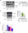
Figure 1. HSV-1 alters APP processing in neuronal cells promoting the formation of 35- and 45-kDa fragments.
(A) Luciferase assay in HeLa cells that had been stably transfected with an APP-Gal4 fusion protein, transiently co-transfected with the G5B-luciferase vector, and infected with HSV-1 (m.o.i. 1). Luciferase activity was measured as an index of APP cleavage at different times p.i.. Data are shown as ratios of values measured in infected cell lysates (INF) to those in control cell lysates (CTR). Each bar represents the mean ratio ± S.D. (n = 6) of 3 individual experiments, each performed in duplicate. *p<0.05 and **p<0.01 vs. 0 h p.i.. (B) Western blot analysis of APP processing in SH-SY5Y cells infected with HSV-1 at different m.o.i. (1, 5, and 10) and harvested 18 h p.i. Blots were probed with 4G8 antibody. The membrane was then stripped and reprobed with anti-actin antibody. Bands representing full-length APPs and APP-F35 are indicated. (C) Western blot analysis of the time course of APP processing in SH-SY5Y cells after HSV-1 infection (m.o.i. 1). The membrane was probed with M2° and then stripped and reprobed with 4G8. Tubulin was used as a loading control. Arrows show bands representing APP-F35 and another APP fragment weighing 45 kDa (APP-F45). (D) Rat cortical neurons were infected and subjected to the same analysis described in C. (E) TCA-precipitated proteins from the supernatants of HSV-1-infected SH-SY5Y cells (showed in figure 1C, 18 h p.i.) were analyzed by western blot with an anti-N-terminal APP antibody (22C11, MAB348) (left panel) and with M2° and 4G8 antibodies (right panels). Released soluble α- and β-APPs (sAPPs), APP-F35 and APP-F45 are indicated. The star in the left panel shows an unidentified 30-kDa N-terminal APP fragment. Results are shown for one representative experiment of three performed.
Figure 2
Figure 2. Host-cell protein synthesis spared by the virus-induced shut-down is necessary for APP-F35 and APP-F45 production.
(A) Real-time PCR assay of APP mRNA levels in SH-SY5Y cells 4, 8 and 18 h after infection with wild-type or mutant (Δγ34.5 and ΔUL41) HSV-1 (m.o.i. 1). APP mRNA levels are expressed as fold changes versus mock-infected cells. At each indicated time point, data are shown as means ± S.D. of 4 independent experiments. For each time *p<0.05 and **p<0.01 vs HSV-1. (B) SH-SY5Y cells were infected with wild-type or mutant (Δγ34.5 and ΔUL41) HSV-1 for 18 h. Extracted proteins were subjected to SDS-PAGE, blotted and probed with M2°, 4G8, and anti-actin antibodies (left). Results are shown for one representative experiment of four performed. Conditioned medium samples were subjected to standard plaque assay to evaluate viral production (right). Data represent the mean ± S.D. of 8 independent experiments, each performed in duplicate.
Figure 3
Figure 3. β- and γ-secretase are involved in APP-F35 formation.
(A) HSV-1-infected (m.o.i. 1) and mock-infected SH-SY5Y cells were treated continuously (1 hour before infection through p.i. hour 18) with 1 µM of a β- or γ-secretase inhibitors (iβ and iγ, respectively). Control cultures (infected and mock-infected) were treated for the same period with equal volumes of solvent (DMSO). Cell lysates were analyzed by western blot with M2° and 4G8 antibodies. Actin was used as loading control. Results are shown for one representative experiment of three performed. Viral production estimated by standard plaque assay is shown next to the western blot. Data are means ± S.D. of 4 independent experiments. (B) Similar experiments were performed independently (as described in A) with 50 µM Z-VAD, which inhibits caspase 3-like enzymes. Densitometric analysis of APP-F35 levels is shown in the graph next to the representative western blot (Z-VAD-treated vs DMSO-treated HSV-1-infected cells) and data are the means ± S.D. of 3 independent experiments performed. Viral production estimated by standard plaque assay is shown. (C) HeLaAG cells were transfected with G5B-Luciferase vector and 24 h later infected with HSV-1 (m.o.i. 1) in the presence of an inhibitor of β-secretase, γ- secretase (1 µM each), or caspase 3-like enzymes (50 µM). Cells were harvested 18 h later and assayed for luciferase activity as a readout of APP cleavage. Data are the means ± S.D. of 3 independent experiments, each performed in duplicate. *p<0.05 and **p<0.01 vs. HSV-1.
Figure 4
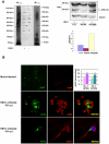
Figure 4. APP-F35 is a soluble oligomer of Aβ peptides.
(A) A pool synthetic Aβ1-42 oligomers and lysates of mock-infected and HSV-1 infected cells were subjected to SDS-PAGE and western blot analysis with 4G8 antibody. A better visualization of the Aβ1-42 oligomeric mixture is provided in the western blot on the right (few seconds of exposure time). The stars indicate the Aβ nonamer whose electrophoretic mobility is similar to that of APP-F35, indicated with the arrow (left panel). HSV-1-infected cells were lysed with 1% triton-X 100, 70% formic acid, or 10% SDS. The samples were resolved by SDS-PAGE, blotted, and immunostained with 4G8 antibody. The membrane was stripped and restained with anti-tubulin antibody (loading control). Western blot is shown for 1 representative experiment of 3 performed (upper right panel). Immunoblots were analyzed densitometrically, and the values were expressed as ratios of APP-F35 to actin (lower right panel). (B) Confocal microscopic images of SH-SY5Y cells 18 h after infection with HSV-1 (m.o.i. 1). Cells were double-labeled with anti-Aβ1-40 and anti-Aβ1-42 antibodies (middle panels) or with anti-Aβ1-42 and anti-HSV-1 antibodies (lower panels). The color of the fluorescence representing each primary antibody is indicated. Results are shown for one representative experiment of three performed. Quantitation of Aβ1-42 from mock- and HSV-1-infected APP695-transfected SH-SY5Y cells by ELISA is shown (upper right). Bar graphs represent the levels (fentomol/mg) of intracellular (pellet) and secreted (medium) Aβ1-42. Data are the means ± S.D. of 3 independent experiments, each performed in duplicate. * p<0.05 vs. HSV-1.
Figure 5
Figure 5. HSV-1 infection promotes nuclear localization of AICD.
Confocal microscopic images of SH-SY5Y cells 18 h after mock or HSV-1 (m.o.i. 1) infection. Cells were labeled with anti-C-terminus-APP antibody (MAB343) and subjected to nuclear DAPI staining. Results are shown for one representative experiment of three performed. Bar graphs showing mean nuclear C-terminus APP labeling intensities in mock- and HSV-1-infected cells. **p<0.01 vs HSV-1 (n = 60). Nuclear and cytoplasmic extracts from mock- and HSV-1-infected cells were immunoprecipitated with anti-APP C-terminus antibody (MAB343) and the samples were resolved by SDS-PAGE. Western blot is one representative experiment of three performed.

References

    1. Schmutzhard E. Viral infections of the CNS with special emphasis on herpes simplex infections. J Neurol. 2001;248:469–477. - PMC - PubMed
    1. Jamieson GA, Maitland NJ, Wilcock GK, Craske J, Itzhaki RF. Latent herpes simplex virus type 1 in normal and Alzheimer’s disease brains. J Med Virol. 1991;33:224–227. - PubMed
    1. Itabashi S, Arai H, Matsui T, Higuchi S, Sasaki H. Herpes simplex virus and risk of Alzheimer’s disease. Lancet. 1997;349:1102. - PubMed
    1. Wozniak MA, Shipley SJ, Combrinck M, Wilcock GK, Itzhaki RF. Productive herpes simplex virus in brain of elderly normal subjects and Alzheimer’s disease patients. J Med Virol. 2005;75:300–306. - PubMed
    1. Kastrukoff L, Hamada T, Schumacher U, Long C, Doherty PC, et al. Central nervous system infection and immune response in mice inoculated into the lip with herpes simplex virus type 1. J Neuroimmunol. 1982;2:295–305. - PubMed

Publication types

MeSH terms

Substances