. 2010 Nov 11;6(11):e1001181.
doi: 10.1371/journal.ppat.1001181.

Host cell invasion and virulence mediated by Candida albicans Ssa1

Affiliations

Host cell invasion and virulence mediated by Candida albicans Ssa1

Jianing N Sun et al. PLoS Pathog. .

Abstract

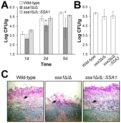
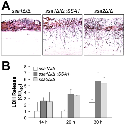
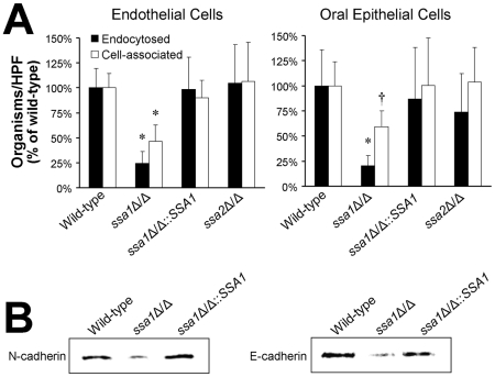
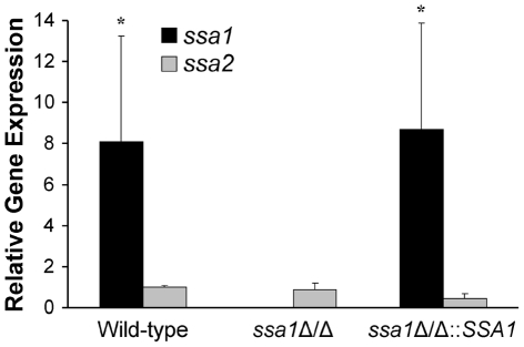
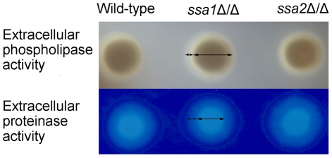
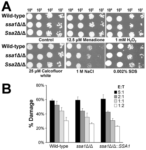
Candida albicans Ssa1 and Ssa2 are members of the HSP70 family of heat shock proteins that are expressed on the cell surface and function as receptors for antimicrobial peptides such as histatins. We investigated the role of Ssa1 and Ssa2 in mediating pathogenic host cell interactions and virulence. A C. albicans ssa1Δ/Δ mutant had attenuated virulence in murine models of disseminated and oropharyngeal candidiasis, whereas an ssa2Δ/Δ mutant did not. In vitro studies revealed that the ssa1Δ/Δ mutant caused markedly less damage to endothelial cells and oral epithelial cell lines. Also, the ssa1Δ/Δ mutant had defective binding to endothelial cell N-cadherin and epithelial cell E-cadherin, receptors that mediate host cell endocytosis of C. albicans. As a result, this mutant had impaired capacity to induce its own endocytosis by endothelial cells and oral epithelial cells. Latex beads coated with recombinant Ssa1 were avidly endocytosed by both endothelial cells and oral epithelial cells, demonstrating that Ssa1 is sufficient to induce host cell endocytosis. These results indicate that Ssa1 is a novel invasin that binds to host cell cadherins, induces host cell endocytosis, and is critical for C. albicans to cause maximal damage to host cells and induce disseminated and oropharyngeal disease.


Conflict of interest statement

SGF holds equity in NovaDigm Therapeutics.

Figures

Figure 1
Figure 1. Ssa1 is required for normal virulence during hematogenously disseminated candidiasis.
Mice were inoculated via the tail vein with 5×10⁵ yeast-phase cells of the indicated strains of C. albicans. (A) Survival over 21 days (n = 10 mice per strain of C. albicans). *P<0.0001 compared to mice infected with either the wild-type or ssa1Δ/Δ::SSA1 complemented strain. (B–D) Fungal burden of the kidneys (B), brains (C), and livers (D) of mice infected with the wild-type, ssa1Δ/Δ and ssa1Δ/Δ::SSA1 complemented strains at the indicated time points. For each organ, the lower bound of the y-axis scale indicates the limits of detection. Results from the 1 day time point are the median ± interquartile range of two experiments with a total of 12 mice per strain of C. albicans. Results from the other time points are the median ± interquartile range of one experiment with 7 mice per strain *P<0.02 compared to mice infected with either the wild-type or ssa1Δ/Δ::SSA1 complemented strain. (E) Periodic acid Schiff stained thin sections of kidneys after 1 day of infection with the indicated strains of C. albicans. Arrows indicate C. albicans filaments in the tissues.
Figure 2
Figure 2. Attenuated virulence of the ssa1Δ/Δ mutant during oropharyngeal candidiasis.
Mice were immunosuppressed with cortisone acetate and then orally inoculated with yeast-phase cells of the indicated strains of C. albicans. After 1, 2, and 5 days of infection, the mice were sacrificed and the tongues and attached tissues were excised. (A) Oral fungal burden of mice infected with the wild-type, ssa1Δ/Δ, and ssa1Δ/Δ::SSA1 complemented strains for the indicated times. Results from the 1 and 2 day time points are the median ± the interquartile range of one experiment with 7 mice per strain of C. albicans. Results from the 5 day time point are the median ± the interquartile range of two experiments with a total of 12 mice per strain. *P≤0.01 compared to both the wild-type and ssa1Δ/Δ::SSA1 strains. (B) Oral fungal burden of mice infected with the wild-type, ssa2, and ssa2Δ/Δ::SSA2 complemented strain for 5 days. Results are the median ± the interquartile range of one experiment with 7 mice per strain of C. albicans. (C) Periodic acid-Schiff stained thin sections of the tongues of mice infected with the indicated strains for 5 days. Arrows indicate the C. albicans filaments.
Figure 3
Figure 3. Ssa1 is necessary for C. albicans to cause maximal damage to endothelial cells and an oral epithelial cell line.
Endothelial cells and the FaDu oral epithelial cell line were incubated with the indicated strains of C. albicans for 3 h, after which the extent of host cell damage was determined using a 51Cr release assay. Results are the mean ± SD of three independent experiments, each performed in triplicate. *P<0.001 compared with the wild-type and ssa1Δ/Δ::SSA1 complemented strains.
Figure 4
Figure 4. The ssa1Δ/Δ mutant has impaired capacity to damage a three-dimensional organotypic model of the oral mucosa.
The three-dimensional model of the oral mucosa was infected by adding 105 yeast-phase cells of the indicated C. albicans strains to the apical surface. (A) Histopathology after 2 days of infection. Thin sections were stained with periodic acid-Schiff. The results are representative of one of three experiments. (B) Epithelial cell damage by the indicated strains was quantified by the release of lactate dehydrogenase (LDH) into the medium. Results are the mean ± SD of 2 experiments.
Figure 5
Figure 5. Ssa1 is necessary for C. albicans to adhere to and be endocytosed by endothelial cells and oral epithelial cells.
(A) Adherence and endocytosis assay. The indicated strains of C. albicans were incubated with endothelial cells or FaDu oral epithelial cells for 90 min, after which the number of endocytosed and cell-associated (endocytosed plus adherent) organisms was determined by a differential fluorescent assay. Results are the mean ± SD of three independent experiments, each performed in triplicate. *P<0.005 compared to the wild-type and ssa1ΔΔ::SSA1 complemented strains; p<0.03 compared to the wild-type and ssa1ΔΔ::SSA1 complemented strains. (B) The capacity of the indicated strains to bind to N-cadherin in extracts of endothelial cell membrane proteins and E-cadherin in extracts of FaDu oral epithelial cell membrane proteins was determined using a whole-cell affinity purification approach. The immunoblot of endothelial cell proteins eluted from hyphae of the indicated strains was developed with an anti-N-cadherin antibody. The immunoblot of epithelial cell proteins eluted from hyphae of the indicated strains was developed with an anti-E-cadherin antibody.
Figure 6
Figure 6. Ssa1 is sufficient to induce endocytosis by host cells.
Latex beads were coated with bovine serum albumin (BSA), recombinant Ssa1 (rSsa1), or rSsa2. They were incubated for 45 min with endothelial cells and FaDu oral epithelial cells, after which the number of endocytosed and cell-associate beads was determined. Data are expressed as a percentage of the results with beads coated with BSA and are the mean ± SD of 3 experiments, each performed in triplicate. *P<0.01 compared to beads coated with BSA.
Figure 7
Figure 7. Ssa1 is located on the cell surface of C. albicans hyphae during epithelial cell interaction.
FaDu oral epithelial cells were infected with C. albicans expressing an Ssa1-GFP fusion protein for 90 min, after which the cells were fixed and stained with an Alexa 594-conjugated anti-C. albicans polyclonal antibody to label the cell surface. (A) Confocal microscopic images of Ssa1-GFP (1) and fluorescent-labeled anti-C. albicans antibody (2). The merged image is shown in (3) and the regions in yellow indicate areas of co-localization between Ssa1-GFP and the fluorescent-labeled anti-C. albicans antibody. (B) Graphs of fluorescent intensity at different cross sections of the hypha in panel (3). The green lines indicate the fluorescent intensity of the Ssa1-GFP and the red lines indicate the fluorescent intensity of the fluorescent-labeled anti-C. albicans antibody. The letters (a-c) correspond to those in panel (3) and indicate the locations along the hypha at which the fluorescent intensity was measured.
Figure 8
Figure 8. Interaction between Ssa1 and Als3.
(A) The ssa1Δ/Δ mutant has normal amounts of Als3 on its surface. Hyphae of the indicated C. albicans strains were stained with a polyclonal anti-Als3 antibody, after which amount of surface exposed Als3 was determined by flow cytometric analysis of 10,000 cells per strain. (B) Normal surface distribution of Als3 on ssa1Δ/Δ hyphae. The distribution of Als3 on the surface of hyphae of the indicated C. albicans strains was determined by staining the organisms with a polyclonal anti-Als3 antibody (green fluorescence) and counterstaining with an anti-C. albicans polyclonal antibody (red fluorescence). (C) Effects of deletion of ALS3 in the ssa1Δ/Δ mutant background. Endothelial cells and FaDu oral epithelial cells were incubated with the indicated strains for 150 min, after which the number of endocytosed and cell-associated organisms were determined. Results are the mean ± SD of 3 experiments, each performed in triplicate. *P<0.01 compared to the wild-type strain, p<0.005 compared to the ssa1Δ/Δ single mutant.
Figure 9
Figure 9. Relative transcript levels of SSA1 and SSA2.
FaDu epithelial cells were infected with the indicated strains of C. albicans for 90 min, after which the C. albicans RNA was extracted. The relative transcript levels of SSA1 and SSA2 were measured by real-time PCR. Results are the mean ± SD of three biological replicates, each tested in duplicate. *P<0.01 compared to SSA2 transcript levels.
Figure 10
Figure 10. C. albicans extracellular phospholipase and protease activities are independent of Ssa1 and Ssa2.
The total extracellular phospholipase activity of the indicated strains was determined by growing them on egg yolk agar at room temperature for 5 days and then measuring the width of the zone of precipitation around each colony (top panel). The total extracellular protease activity of the indicated strains was assessed by growing them on BSA agar at 37°C for 5 days and then staining the plates with 0.5% amido black. The extracellular protease activity was proportional to the width of the zone of clearance around the colonies (lower panel).
Figure 11
Figure 11. Ssa1 and Ssa2 are not required for resistance to stress.
(A) Susceptibility to oxidant and cell wall stress. Serial 10-fold dilutions of the indicated strains were used to inoculate YPD plates containing menadione, H2O2, Calcofluor white, NaCl, or SDS. Images were obtained after growth at 30°C for 24 h. (B) Susceptibility to leukocyte induced damage. Yeast-phase cells of the indicated strains were incubated for 3 h with HL-60 cells that had been differentiated into neutrophil-like cells, after which the amount of damage to the organisms was determined by an XTT assay. Each strain was tested at an effector to target ratio (E:T) ranging from 5∶1 to 1∶2. Results are the mean ± SD of three independent experiments, each performed in triplicate.

References

    1. Wisplinghoff H, Bischoff T, Tallent SM, Seifert H, Wenzel RP, et al. Nosocomial bloodstream infections in US hospitals: analysis of 24,179 cases from a prospective nationwide surveillance study. Clin Infect Dis. 2004;39:309–317. - PubMed
    1. Filler SG. Candida-host cell receptor-ligand interactions. Curr Opin Microbiol. 2006;9:333–339. - PubMed
    1. Grubb SE, Murdoch C, Sudbery PE, Saville SP, Lopez-Ribot JL, et al. Candida albicans-endothelial cell interactions: a key step in the pathogenesis of systemic candidiasis. Infect Immun. 2008;76:4370–4377. - PMC - PubMed
    1. Walker LA, Maccallum DM, Bertram G, Gow NA, Odds FC, et al. Genome-wide analysis of Candida albicans gene expression patterns during infection of the mammalian kidney. Fungal Genet Biol. 2009;46:210–219. - PMC - PubMed
    1. Rodaki A, Bohovych IM, Enjalbert B, Young T, Odds FC, et al. Glucose promotes stress resistance in the fungal pathogen Candida albicans. Mol Biol Cell. 2009;20:4845–4855. - PMC - PubMed

Publication types