. 2010 Nov 11;6(11):e1001186.
doi: 10.1371/journal.ppat.1001186.

Ubiquitin-regulated nuclear-cytoplasmic trafficking of the Nipah virus matrix protein is important for viral budding

Affiliations

Ubiquitin-regulated nuclear-cytoplasmic trafficking of the Nipah virus matrix protein is important for viral budding

Yao E Wang et al. PLoS Pathog. .

Abstract

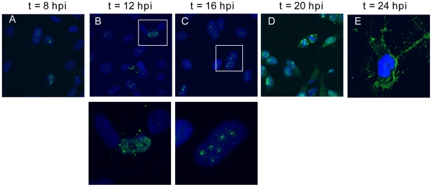
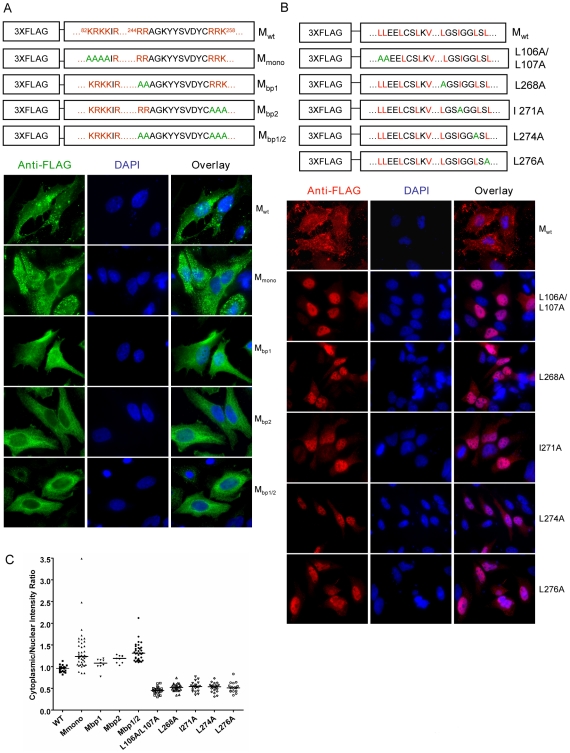
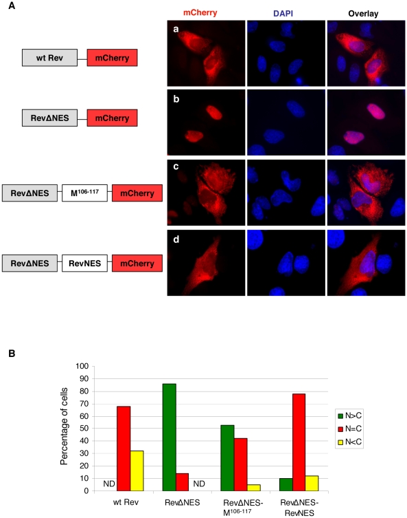
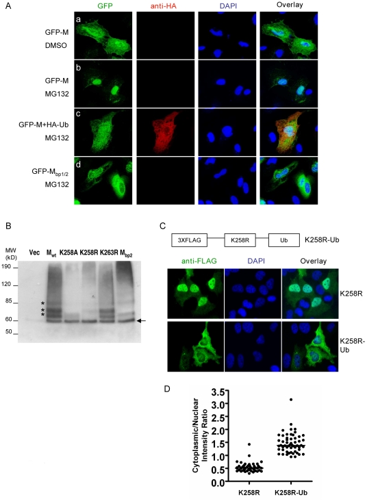
Paramyxoviruses are known to replicate in the cytoplasm and bud from the plasma membrane. Matrix is the major structural protein in paramyxoviruses that mediates viral assembly and budding. Curiously, the matrix proteins of a few paramyxoviruses have been found in the nucleus, although the biological function associated with this nuclear localization remains obscure. We report here that the nuclear-cytoplasmic trafficking of the Nipah virus matrix (NiV-M) protein and associated post-translational modification play a critical role in matrix-mediated virus budding. Nipah virus (NiV) is a highly pathogenic emerging paramyxovirus that causes fatal encephalitis in humans, and is classified as a Biosafety Level 4 (BSL4) pathogen. During live NiV infection, NiV-M was first detected in the nucleus at early stages of infection before subsequent localization to the cytoplasm and the plasma membrane. Mutations in the putative bipartite nuclear localization signal (NLS) and the leucine-rich nuclear export signal (NES) found in NiV-M impaired its nuclear-cytoplasmic trafficking and also abolished NiV-M budding. A highly conserved lysine residue in the NLS served dual functions: its positive charge was important for mediating nuclear import, and it was also a potential site for monoubiquitination which regulates nuclear export of the protein. Concordantly, overexpression of ubiquitin enhanced NiV-M budding whereas depletion of free ubiquitin in the cell (via proteasome inhibitors) resulted in nuclear retention of NiV-M and blocked viral budding. Live Nipah virus budding was exquisitely sensitive to proteasome inhibitors: bortezomib, an FDA-approved proteasome inhibitor for treating multiple myeloma, reduced viral titers with an IC(50) of 2.7 nM, which is 100-fold less than the peak plasma concentration that can be achieved in humans. This opens up the possibility of using an "off-the-shelf" therapeutic against acute NiV infection.


Conflict of interest statement

The authors have declared that no competing interests exist.

Figures

Figure 1
Figure 1. Nuclear-cytoplasmic trafficking of Nipah virus matrix protein (NiV-M) during live viral infection.
HeLa cells plated on poly-lysine-coated glass coverslips were incubated with Nipah virus Malaysia strain for 1 hr at 37°C and then fresh growth medium for up to 24 hrs. At (A) 8, (B) 12, (C) 16, (D) 20, and (E) 24 hpi, cells were fixed with 10% formalin, stained with rabbit anti-M polyclonal antibody and imaged on a confocal fluorescent microscope (63× magnification). DAPI was used for visualization of the nuclei. Insets in (B) and (C) indicate nuclear localization of M in infected cells. Experiments were performed under BSL4 conditions.
Figure 2
Figure 2. Mutagenesis studies of potential nuclear localization signals (NLSs) and nuclear export signals (NESs) in NiV-M.
Positively charged amino acid residues in the predicted monopartite and bipartite NLSs (A) or key leucine/isoleucine residues in the potential NESs (B) were mutated to alanines using site-directed mutagenesis. HeLa cells expressing the indicated proteins were stained with an anti-FLAG monoclonal antibody as well as DAPI. Representative fields are shown in (A) and (B), and (C) shows the quantification of cytoplasmic/nuclear fluorescence intensity (C:N) ratios for ∼10–50 individual cells analyzed for each mutant as described in Materials and Methods . Compared to Mwt, statistically significant increases in C:N ratios were observed for Mbp1 (p<0.01), Mbp2 (p<0.0001) and Mbp1/2 (p<0.0001) (unpaired t-test).
Figure 3
Figure 3. NiV-M NES partially restores nuclear export to an NES-defective HIV Rev.
(A) HeLa cells were transiently transfected with plasmids encoding Rev-mCherry (panel a), RevΔNES-mCherry (panel b), RevΔNES-M106–117-mCherry (panel c), or RevΔNES-RevNES-mCherry (panel d). 24 hrs post transfection, cells were treated with 5 µg/ml actinomycin D for 4 hrs before fixation. Cells were stained with DAPI for visualization of the nuclei and imaged on a fluorescent microscope under 60× magnification. Representative images are shown in (A), and (B) shows the quantification of the percentage of cells with the fusion protein localized to only the nucleus (N>C), both the nucleus and the cytoplasm (N = C), or only the cytoplasm (N<C). For each mutant, at least 100 cells were counted. Both M106–117 and the endogenous NES from Rev were able to restore nuclear export to the RevΔNES-mCherry fusion protein.
Figure 4
Figure 4. Correlation between the nuclear localization of NiV-M and VLP budding.
Viral-like particles were harvested from culture supernatants of cells expressing wild-type NiV-M, NLS mutants (A) or NES mutants (B) at 24 hpt as described in Materials and Methods . VLPs and the corresponding cell lysates were immunoblotted with an anti-FLAG antibody. The cell lysate blots were then stripped and re-probed with an anti-β-tubulin antibody as loading control. Representative results are shown in (A) and (B). (C) and (D) show the quantification of the budding index for the indicated wild-type and mutant NiV-M proteins as described in Materials and Methods . Error bars were calculated from three independent experiments. M mutants that were deficient in either nuclear import or export were also deficient in budding.
Figure 5
Figure 5. Dual functions of critical residue K258 in regulating NiV-M nuclear-cytoplasmic trafficking.
(A) The matrix protein sequences of twelve viruses from different genera within the family Paramyxoviridae were aligned using CLUSTAL W (version 1.83). Positively charged amino acid residues that conform to the consensus for bipartite NLSs are colored green. The red arrow points to the lysine residue conserved among all twelve viruses. (B) K258 in NiV-M was mutated to alanine or arginine using site-directed mutagenesis. As control, K263, a non-conserved lysine in the vicinity of K258, was also mutated to arginine. HeLa cells transfected with the indicated constructs were stained with mouse anti-FLAG antibody and DAPI. K258A was excluded from the nucleus, whereas K258R was concentrated in the nucleus. The localization of K263R was similar to wild-type M. Quantification of cytoplasmic/nuclear fluorescence intensity ratios is shown in (C).
Figure 6
Figure 6. Ubiquitination regulates NiV-M nuclear export.
(A) Ubiquitin depletion by MG132 treatment inhibits M nuclear export. HeLa cells were transfected with GFP-M alone (panels a and b), GFP-M plus HA-Ub (panel c) or GFP-Mbp1/2 (panel d). 24 hpt, cells were treated with 50 µM MG132 or DMSO as indicated, fixed 6 hrs later with 2% paraformaldehyde, stained with DAPI as well as a mouse anti-HA antibody followed by Alexa594-conjugated goat-anti-mouse secondary antibody to identify cells expressing HA-Ub, and imaged on a fluorescent microscope. Representative images are shown. (B) Ubiquitination patterns of wild-type M and the indicated mutants. HEK293T cells were co-transfected with HA-Ub (in which all the lysines were mutated to arginines to specifically look at monoubiquitination) and the indicated 3XFLAG-tagged M mutants or empty vector as control. M was immunoprecipitated as described in Materials and Methods and the ubiquitinated species were detected by immunoblotting using an anti-HA antibody. The banding patterns of K258A, K258R and Mbp2 were different from Mwt, whereas K263R was similar to Mwt. (C) Mimicking monoubiquitination restores nuclear export to K258R. One copy of ubiquitin was fused in frame to the C-terminus of 3XFLAG-K258R, and HeLa cells expressing K258R or K258R-Ub were stained with an anti-FLAG antibody. Quantification of the cytoplasmic/nuclear fluorescence intensity ratio for each mutant is shown in (D). There is significant difference between the localization patterns of K258R and K258R-Ub (p<0.0001, unpaired t test).
Figure 7
Figure 7. K258 is critical for NiV-M membrane association and budding.
(A) NiV-M K258 mutants are deficient in VLP budding. VLP and cell lysate samples were prepared from cells expressing wild-type M, K258A, K258R or K263R at 24 hpt as described in Materials and Methods . Immunoblotting was performed using an anti-FLAG monoclonal antibody, then the cell lysate blot was stripped and re-probed with an anti-β-tubulin antibody as loading control. Both K258A and K258R were expressed in the cells at similar levels compared to wild-type M, but they were absent from the VLPs. The experiment was repeated three times and representative results are shown. (B) Quantification of the budding index for the wild-type and mutant NiV-M proteins shown in (A). (C) Wild-type M localized to membrane patches and fine filopodia extensions while the K258A mutant did not. (D) K258A is deficient in membrane association. HEK293T cells expressing wild-type NiV-M, K258A, wild-type HIV Gag, or a myristoylation mutant of HIV Gag (G2A) were harvested at 24 hpt. Cell homogenates were loaded at the bottom of a 10–73% discontinuous sucrose gradient and ultracentrifuged for 16 hrs at 100,000×g. Eight fractions were collected from the top, and proteins were extracted using methanol/chloroform prior to immunoblotting with anti-NiV-M (in the case of Mwt and K258A) or anti-myc (Gagwt and G2A) antibodies. Membrane-associated proteins were collected at the interface between 10% and 65% sucrose as “fraction 2” as described previously . (E) Fusion to L10 or S15, the membrane targeting N-terminal peptide sequence from p56lck and c-Src, respectively, restores membrane association to the K258A mutant. Membrane flotation centrifugation was performed as in (D). (F) Rescue of K258A budding by L10 and S15. VLP and cell lysate samples were prepared from HEK293T cells expressing the indicated constructs and examined by immunoblotting using a rabbit anti-NiV-M antibody. The cell lysate blot was also probed with an anti-β-tubulin antibody as loading control. The experiment was repeated three times. Representative blots are shown in (F), and the quantification of the budding indices is shown in (G).
Figure 8
Figure 8. MG132 and bortezomib inhibit NiV-M nuclear export during live viral infection and reduce viral titers.
(A) NiV-M VLP budding in the presence of MG132. HEK293T cells expressing 3XFLAG-M (left three lanes) or 3XFLAG-M plus HA-Ub (right two lanes) were incubated with DMSO, 10 µM or 50 µM MG132 for 12 hrs, and VLPs produced during this period were harvested as described in Materials and Methods . VLPs and cell lysates were immunoblotted with an anti-FLAG antibody, then the cell lysate blot was stripped and re-probed with an anti-β-tubulin antibody as loading control. (B) MG132 altered M localization during live viral infection. HeLa cells infected with Nipah virus Malaysia strain were incubated with 50 µM MG132 or DMSO for 8 hrs starting from 15 hpi. Cells were then stained with an anti-M antibody and imaged on a confocal microscope. MG132 restricted M localization to the nuclear compartment. (C) and (D) Dose-response curves of Nipah viral titers in the presence of MG132 (C) or bortezomib (D). HeLa cells were incubated with NiV for 1 hr at 37°C and then fresh growth medium. 15 hpi, serial dilutions of MG132 or bortezomib were added, yielding final concentrations ranging from 10 nM to 1 fM. Considering the short half-life of bortezomib (9–15 hrs), it was re-added 12 hrs later. Supernatants were collected at 40 hpi and viral titers were determined by plaque assay. To calculate the 50% inhibitory concentration (IC50), the resulting data were fit to the sigmoidal dose-response curve (GraphPad Prism software version 4.00) using the equation: % inhibition =  minimal inhibition + (maximal inhibition-minimal inhibition)/(1+10∧(LogIC50-Log drug concentration)). Results shown are from two independent experiments with triplicates for each data point. The infected cells were harvested, and the expression of cellular (β-actin) and viral (matrix) proteins was examined by immunoblotting.

Similar articles

Cited by

References

    1. Eaton BT, Broder CC, Middleton D, Wang LF. Hendra and Nipah viruses: different and dangerous. Nat Rev Microbiol. 2006;4:23–35. - PMC - PubMed
    1. Field H, Young P, Yob JM, Mills J, Hall L, et al. The natural history of Hendra and Nipah viruses. Microbes Infect. 2001;3:307–314. - PubMed
    1. Chua KB, Bellini WJ, Rota PA, Harcourt BH, Tamin A, et al. Nipah virus: a recently emergent deadly paramyxovirus. Science. 2000;288:1432–1435. - PubMed
    1. Weingartl HM, Berhane Y, Czub M. Animal models of henipavirus infection: a review. Vet J. 2009;181:211–220. - PubMed
    1. Hsu VP, Hossain MJ, Parashar UD, Ali MM, Ksiazek TG, et al. Nipah virus encephalitis reemergence, Bangladesh. Emerg Infect Dis. 2004;10:2082–2087. - PMC - PubMed

Publication types

MeSH terms