. 2010 Nov 11;6(11):e1000994.
doi: 10.1371/journal.pcbi.1000994.

The role of oligomerization and cooperative regulation in protein function: the case of tryptophan synthase

Affiliations

The role of oligomerization and cooperative regulation in protein function: the case of tryptophan synthase

M Qaiser Fatmi et al. PLoS Comput Biol. .

Abstract

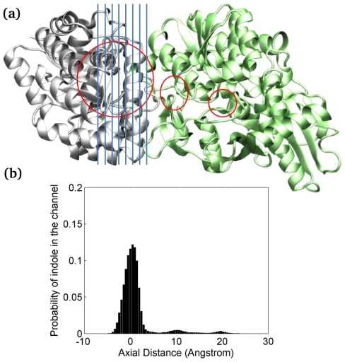
The oligomerization/co-localization of protein complexes and their cooperative regulation in protein function is a key feature in many biological systems. The synergistic regulation in different subunits often enhances the functional properties of the multi-enzyme complex. The present study used molecular dynamics and Brownian dynamics simulations to study the effects of allostery, oligomerization and intermediate channeling on enhancing the protein function of tryptophan synthase (TRPS). TRPS uses a set of α/β-dimeric units to catalyze the last two steps of L-tryptophan biosynthesis, and the rate is remarkably slower in the isolated monomers. Our work shows that without their binding partner, the isolated monomers are stable and more rigid. The substrates can form fairly stable interactions with the protein in both forms when the protein reaches the final ligand-bound conformations. Our simulations also revealed that the α/β-dimeric unit stabilizes the substrate-protein conformation in the ligand binding process, which lowers the conformation transition barrier and helps the protein conformations shift from an open/inactive form to a closed/active form. Brownian dynamics simulations with a coarse-grained model illustrate how protein conformations affect substrate channeling. The results highlight the complex roles of protein oligomerization and the fine balance between rigidity and dynamics in protein function.


Conflict of interest statement

The authors have declared that no competing interests exist.

Figures

Figure 1
Figure 1. Structure of the α/β–dimeric unit of tryptophan synthase.
(a) A labeled diagram of the α/β–dimeric unit of tryptophan synthase (TRPS). The important regions have been color coded: blue for α–L2 (residues α53–60), red for α–L6 (residues α179–193), purple+pink for the β–COMM domain (residues β102–189), and pink for the β–H6 of COMM domain (residues β165–181). The approximate location of the interconnecting channel is shown as a solid brown line. (b) The network of H–bonds at the α/β–interface of the TRPS dimeric unit. Some of the H–bonds play key roles in allosteric communications and the substrate channeling process. The interacting pair of residues is zoomed in, and the formation of possible H–bonds is shown in small panels.
Figure 2
Figure 2. H–bond formations in different states.
A quantitative comparison of H–bond formations in percentages for residues located at the α/β–interface of ligand–free (LF; blue bars) and ligand–bound–reference (LBR; red bars) dimeric units obtained from the molecular dynamics (MD) simulations of the TRPS complex. The formation of the H–bonds between α– and β–interfacial residues is given in (a), while (b) shows the formation of the H–bonds of the α– and β–interfacial residues within α– and β–subunits, respectively. The x–axis is labeled with the interacting pair of residues; the α–residues are labeled green and the β–residues are black.
Figure 3
Figure 3. Comparison of correlated motions between different states.
The comparison of correlated motions in the dimeric units with (a) ligand–free and (b) ligand–bound–reference states. The labeled α–residues on the x– and y–axes have a white background and the β–residues have a black background. Red, blue and pink rectangular boxes represent residues in α–L2, α–L6 and β–H6 of the COMM domain, respectively. The interfacial residues in both α– and β–subunits are underlined in green.
Figure 4
Figure 4. Plots of RMSFs.
Comparative root mean square fluctuations (RMSFs) for the α–subunits in the dimeric complexes versus isolated α–monomeric units based on Cα atoms for each residue, averaged over the total simulation time of 50 ns of the ligand–free (LF) and ligand–bound–reference (LBR) states.
Figure 5
Figure 5. H–bond network in the dimeric and monomeric complexes.
Quantitative comparison of the H–bond network for the dimeric and monomeric complexes of the ligand–bound–reference (LBR) state for residues located at the interface and residues interacting with ligands in the active sites. (a) α–subunit in the α/β–dimeric unit versus α–monomeric unit, (b) β–subunit in the α/β–dimeric unit versus β–monomeric unit. IGP and A-A are the α– and β–site ligands and represent 3-indole-D-glycerol-3′-phosphate and aminoacrylate, respectively. The x–axis is labeled with the interacting pair of residues, with the ligands blue and the protein residues in black.
Figure 6
Figure 6. Comparison of correlated motions between the dimeric and monomeric complexes.
Comparison of correlated motions between the α–subunit in the α/β–dimeric unit (a) versus the α–monomeric unit (b) and the β–subunit in the α/β–dimeric unit (c) versus the β–monomeric unit (d) for the ligand–bound–reference (LBR) state, calculated with MutInf. Red, blue and pink rectangular boxes represent residues in α–L2, α–L6 and β–H6 of COMM domain, respectively. The interfacial residues are underlined in green and the residues at the active sites are underlined in cyan.
Figure 7
Figure 7. Brownian dynamics simulations in different states.
Snapshots taken from the Brownian dynamics simulations of ligand–free (a), ligand–bound (b), and ligand–bound–reference (c) states of the α/β–dimeric units showing the leakage, blockage and passage of the indole intermediate, respectively, during the channeling process. Indole is represented by one yellow bead.
Figure 8
Figure 8. Analysis of indole distribution.
Analysis of indole distribution in the channel from 360 individual coarse-grained Brownian dynamics (CGBD) simulations. The protein conformation is taken from a 30-ns MD simulation in the LBR state. (a) The channel is divided into 72 sections through the α–active site to the β–active site. Regions that indole spent most of the time during transportation are circled in red. The blue vertical lines shown in the plot does not reflect the real size of each section. (b) A histogram indicates regions of channel where indole preferentially resides.

Similar articles

Cited by

References

    1. Woolf PJ, Linderman JJ. Self organization of membrane proteins via dimerization. Biophys Chem. 2003;104:217–227. - PubMed
    1. Marianayagam NJ, Sunde M, Matthews JM. The power of two: protein dimerization in biology. Trends Biochem Sci. 2004;29:618–625. - PubMed
    1. Meng GY, Fronzes R, Chandran V, Remaut H, Waksman G. Protein oligomerization in the bacterial outer membrane. Mol Membr Biol. 2009;26:136–145. - PubMed
    1. Ali MH, Imperiali B. Protein oligomerization: How and why. Bioorgan Med Chem. 2005;13:5013–5020. - PubMed
    1. Brinda KV, Vishveshwara S. Oligomeric protein structure networks: insights into protein-protein interactions. BMC Bioinformatics. 2005;6 doi: 10.1186/1471-2105-6-296. - DOI - PMC - PubMed

Publication types