

Functional analysis of Ficolin-3 mediated complement activation
- PMID: 21085669
- PMCID: PMC2978102
- DOI: 10.1371/journal.pone.0015443
Functional analysis of Ficolin-3 mediated complement activation
Abstract
The recognition molecules of the lectin complement pathway are mannose-binding lectin and Ficolin -1, -2 and -3. Recently deficiency of Ficolin-3 was found to be associated with life threatening infections. Thus, we aimed to develop a functional method based on the ELISA platform for evaluating Ficolin-3 mediated complement activation that could be applicable for research and clinical use. Bovine serum albumin (BSA) was acetylated (acBSA) and chosen as a solid phase ligand for Ficolins in microtiter wells. Binding of Ficolins on acBSA was evaluated, as was functional complement activation assessed by C4, C3 and terminal complement complex (TCC) deposition. Serum Ficolin-3 bound to acBSA in a calcium dependent manner, while only minimal binding of Ficolin-2 and no binding of Ficolin-1 were observed. No binding to normal BSA was seen for any of the Ficolins. Serum C4, C3 and TCC deposition on acBSA were dependent only on Ficolin-3 in appropriate serum dilutions. Deposition of down stream complement components correlated highly significantly with the serum concentration of Ficolin-3 but not with Ficolin-2 in healthy donors. To make the assay robust for clinical use a chemical compound was applied to the samples that inhibited interference from the classical pathway due to the presence of anti-BSA antibodies in some sera. We describe a novel functional method for measuring complement activation mediated by Ficolin-3 in human serum up to the formation of TCC. The assay provides the possibility to diagnose functional and genetic defects of Ficolin-3 and down stream components in the lectin complement pathway.
Conflict of interest statement
Figures
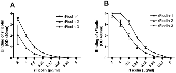
 ), rFicolin-2 (
 ), rFicolin-3 (
 ) at (A) 30 min at 37°C or (B) 3 h at RT. Binding was detected with monoclonal antibodies directed against the individual proteins. Graphs show mean ± SD (n = 4).
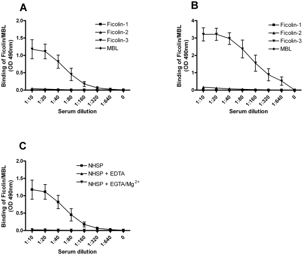
 ), Ficolin-2 (
 ), Ficolin-3 (
 ) or MBL (
 ) was subsequently detected with monoclonal antibodies directed against the individual proteins. (C) acBSA incubated with: NHSP (
 ), NHSP with 10 mM EDTA (
 ) or NHSP with 10 mM EGTA and 5 mM Mg2+ (
 ). Binding was detected with a monoclonal antibody against Ficolin-3. All graphs show mean ± SD (n = 3).
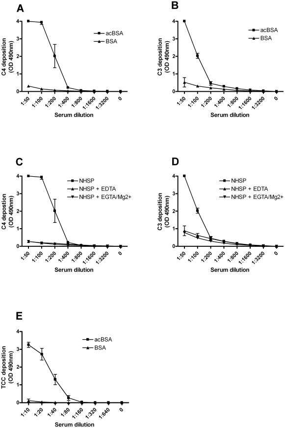
 ) or BSA (
 ) was detected using a polyclonal antibody. (C–D) Microtiter wells were coated with acBSA and subsequently incubated with NHSP (
 ), NHSP with 10 mM EDTA (
 ) or NHSP with 10 mM EGTA and 5 mM Mg2+ (
 ) for 30 min at 37°C. (C) C4 or (D) C3 deposition was detected as described above. (E) Finally, microtiter wells were coated with acBSA (
 ) or BSA (
 ) and subsequently incubated with NHSP in serial dilution for 45 min at 37°C and TCC-deposition on acBSA was detected using a monoclonal antibody. All graphs show mean ± SD (n = 4).
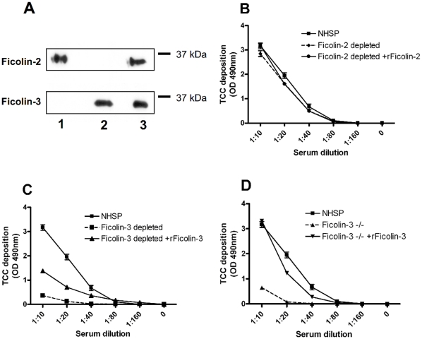
 ), Ficolin-2 depleted serum (-♦-), Ficolin-2 depleted serum with addition of 5 µg/ml rFicolin-2 (
 ). (C) Shows TCC deposition for: NHSP (
 ), Ficolin-3 depleted serum (-▪-) and Ficolin-3 depleted serum with addition of 25 µg/ml rFicolin-3 (
 ). (D) Shows TCC deposition for: NHSP (
 ), Ficolin-3 deficient serum −/− (-▴-), Ficolin-3 deficient serum −/− with addition of 25 µg/ml rFicolin-3 (
 ). All graphs show mean ± SD of duplicate wells.
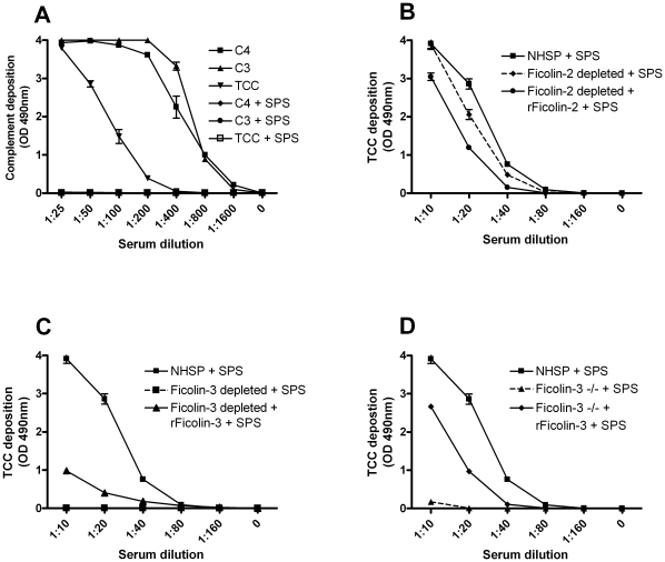
 ), C3 (
 ) or TCC (
 ); and in the presence of SPS: C4 (
 ), C3 (-•-) or TCC (-□-). (B–D) Different sera pre-incubated on ice with SPS and then incubated for 45 min at 37°C in microtiter plates coated with acBSA before TCC deposition was detected with a monoclonal antibody. (B) Shows TCC deposition in the presence of SPS for: NHSP (
 ), Ficolin-2 depleted serum (- ♦ -), Ficolin-2 depleted serum with addition of 5 µg/ml rFicolin-2 (
 ). (C) Shows TCC deposition in the presence of SPS for: NHSP (
 ), Ficolin-3 depleted serum (-▪-) and Ficolin-3 depleted serum with addition of 25 µg/ml rFicolin-3 (
 ). (D) Shows TCC deposition in the presence of SPS for: NHSP (-▪-), Ficolin-3 deficient serum −/− (-▴-), Ficolin-3 deficient serum −/− with addition of 25 µg/ml rFicolin-3 (
 ). All graphs show mean ± SD of duplicate wells.
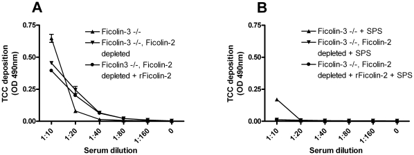
 ), Ficolin-3 deficient serum −/− depleted of Ficolin-2 (
 ), Ficolin-3 deficient serum −/− depleted of Ficolin-2 with addition of 5 µg/ml rFicolin-2 (
 ). TCC deposition was detected with a monoclonal antibody. Graphs show mean ± SD of duplicate wells.
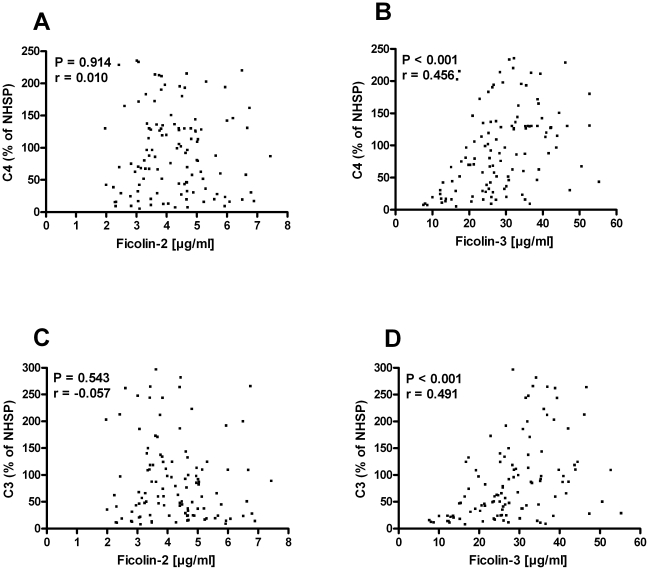
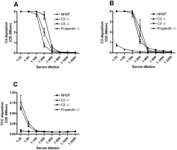
 ), C2 deficient serum (
 ), C5 deficient serum (
 ) or properdin deficient serum (
 ) in serial dilutions for 30 min at 37°C. (A) C4 deposition and (B) C3 deposition on acBSA measured using polyclonal antibodies. (C) TCC deposition on acBSA measured using a monoclonal antibody. All graphs show mean ± SD (n = 3).
References
-
- Walport MJ. Complement. First of two parts. N Engl J Med. 2001;344:1058–1066. - PubMed
-
- Kishore U, Reid KB. C1q: structure, function, and receptors. Immunopharmacology. 2000;49:159–170. - PubMed
-
- Spitzer D, Mitchell LM, Atkinson JP, Hourcade DE. Properdin can initiate complement activation by binding specific target surfaces and providing a platform for de novo convertase assembly. J Immunol. 2007;179:2600–2608. - PubMed
-
- Thiel S. Complement activating soluble pattern recognition molecules with collagen-like regions, mannan-binding lectin, ficolins and associated proteins. Mol Immunol. 2007;44:3875–3888. - PubMed
Publication types
MeSH terms
Substances
LinkOut - more resources
Full Text Sources
Other Literature Sources
Molecular Biology Databases
Miscellaneous
