. 2010 Nov 11;5(11):e13921.
doi: 10.1371/journal.pone.0013921.

α-actinin-4 is essential for maintaining the spreading, motility and contractility of fibroblasts

Affiliations

α-actinin-4 is essential for maintaining the spreading, motility and contractility of fibroblasts

Hanshuang Shao et al. PLoS One. .

Abstract

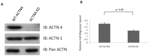
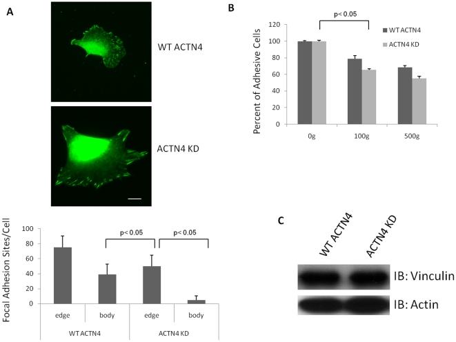
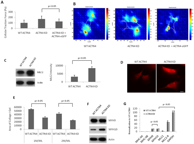
Background: α-Actinins cross-link actin filaments, with this cross-linking activity regulating the formation of focal adhesions, intracellular tension, and cell migration. Most non-muscle cells such as fibroblasts express two isoforms, α-actinin-1 (ACTN1) and α-actinin-4 (ACTN4). The high homology between these two isoforms would suggest redundancy of their function, but recent studies have suggested different regulatory roles. Interestingly, ACTN4 is phosphorylated upon growth factor stimulation, and this loosens its interaction with actin.

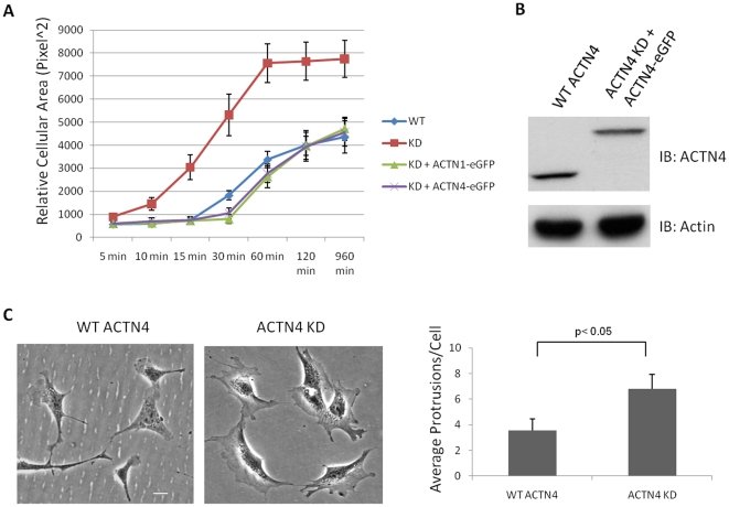
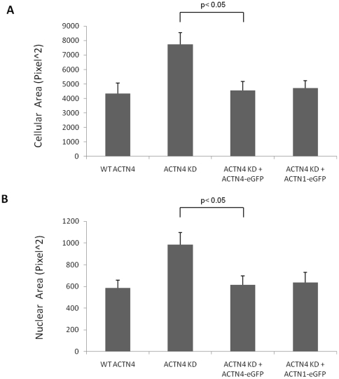
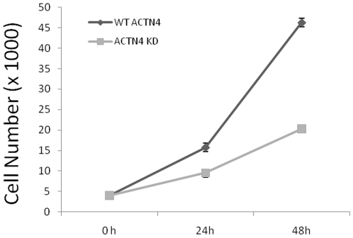
Methodology/principal findings: Using molecular, biochemical and cellular techniques, we probed the cellular functions of ACTN4 in fibroblasts. Knockdown of ACTN4 expression in murine lung fibroblasts significantly impaired cell migration, spreading, adhesion, and proliferation. Surprisingly, knockdown of ACTN4 enhanced cellular compaction and contraction force, and increased cellular and nuclear cross-sectional area. These results, except the increased contractility, are consistent with a putative role of ACTN4 in cytokinesis. For the transcellular tension, knockdown of ACTN4 significantly increased the expression of myosin light chain 2, a element of the contractility machinery. Re-expression of wild type human ACTN4 in ACTN4 knockdown murine lung fibroblasts reverted cell spreading, cellular and nuclear cross-sectional area, and contractility back towards baseline, demonstrating that the defect was due to absence of ACTN4.

Significance: These results suggest that ACTN4 is essential for maintaining normal spreading, motility, cellular and nuclear cross-sectional area, and contractility of murine lung fibroblasts by maintaining the balance between transcellular contractility and cell-substratum adhesion.


Conflict of interest statement

Competing Interests: The authors have declared that no competing interests exist.

Figures

Figure 1
Figure 1. Knockdown of ACTN4 impairs cell migration of murine lung fibroblasts.
(A) WT ACTN4 and ACTN4 KD murine lung fibroblasts grown in 6-well Petri-dish plates were lysed with RIPA buffer in the presence of 1× proteinase cocktail mix. Equal amount of total protein were separated by 7.5% SDS-PAGE and immunoblotted with anti-ACTN4, ACTN1 and pan-ACTN. The data are representative of three independent experiments. (B) Cells were plated on 6-well plastic dishes and grown to confluence in DMEM in the presence 10% fetal bovine serum. A scraped area was made by a rubber policeman and then cultured for additional 24 hr. Photographs were taken at 0 hr and 24 hr, and the relative distance migrated by the cells was determined by using Photoshop software. Data are mean ± s.e.m., measured from three independent experiments each in triplicate.
Figure 2
Figure 2. Knockdown of ACTN4 hastens cell spreading of murine lung fibroblasts.
(A) Murine lung fibroblasts (WT ACTN4 and ACTN4 KD) were transiently transfected to express the designated constructs. These cells were trypsined and then plated in 6-well plates coated with 1µg/ml fibronectin. After culturing for 5 min, 10 min, 15 min, 30 min, 1 hr, 2 hr and 16 hr, respectively, unattached cells were washed away with PBS. Adhesive cells were fixed with 2% formaldehyde for 30 min at room temperature followed by imaging under fluorescent microscope. The cellular cross-sectional area of attached cells with GFP fluorescence was measured by using ImageJ software. Quantification represents the average cellular cross-sectional area (± the standard deviation) of at least 50 individual fluorescent cells chosen randomly. (B) Immunoblotting of endogenous ACTN4 and exogenous human wild type ACTN4-eGFP. The immunoblotting results were the representative of three independent experiments. (C) Morphology of both WT ACTN4 and ACTN4 KD fibroblasts grown in Petri dishes and the quantification of protrusions per cell. Images present both WT and KD fibroblasts chosen randomly. Quantification represents the average protrusions per cell (± the standard deviation) of at least 50 individual cells chosen randomly. Bar  = 5 µm.
Figure 3
Figure 3. Knockdown of ACTN4 limits the number of focal adhesions.
(A) WT ACTN4 and ACTN4 KD murine lung fibroblasts were stained for vinculin aggregates as indicative of focal adhesions. The number of focal adhesion sites residing within cell body and cellular edge were calculated by using ImageJ software for at least 30 individual cells. The ratio of focal adhesion sites between cell body and edge was also calculated. Images were taken randomly. Quantification was measured from at least 30 individual cells (± the standard deviation) chosen randomly. Shown are representative of at least independent experiments. Bar  = 10 µm. (B) An inverted centrifugation focal adhesion assay was performed (see “material and method” for details). Data are mean ± s.e.m., measured from three independent experiments. (C) Immunoblotting shows that the vinculin level was similar in WT and KD cells. Blot is representative of three independent assessments.
Figure 4
Figure 4. Knockdown of ACTN4 increases both cellular and nuclear cross-sectional area of murine lung fibroblasts.
Murine lung fibroblasts of WT ACTN4 and ACTN4 KD transiently expressed eGFP and ACTN4 KD transiently expressed ACTN4-eGFP or ACTN1-eGFP were trypsined and then plated in 6-well Petri-dish plates coated with 1 µg/ml fibronectin. After incubation for 16 hr, cells were further incubated with 10 µg/ml Hoechst 33342 for 10 min at 37°C in 5% CO2 incubator prior to fixation with 2% formaldehyde for 30 min at room temperature. After taking fluorescent images under microscope, the cellular cross-sectional area of attached cells with GFP fluorescence (A) and the nuclear cross-sectional area with blue fluorescence (B) were measured by using ImageJ software. Quantification represents the average cellular and nuclear cross-sectional area (± the standard deviation) of at least 50 individual cells chosen randomly.
Figure 5
Figure 5. Knockdown of ACTN4 increases both contractile force and gel compaction generated by murine lung fibroblasts.
(A) Contractile force assays were performed with both WT ACTN4 and ACTN4 KD murine lung fibroblasts transiently transfected with either eGFP or human wild type ACTN4-eGFP. For each cell type, at least 20 individual cells with GFP fluorescence were analyzed. Quantification represents the average traction force per cell (± the standard deviation) of at least 20 individual fluorescent cells chosen randomly. Shown are representative of at least independent experiments. (B) Images created by using special software. The length of arrows stands for the strength of traction force. Shown are representative images of three independent experiments. (C) Immunoblotting of MLC2 of total cellular lysate and actin as loading control. The bands of MLC2 were quantitated by using ImageJ software. Shown are representative blot of three independent experiments. Data are mean ±., measured from three independent experiments. (D) Immunofluorescence of MLC2 in both ACTN4 and ACTN4 KD murine lung fibroblasts. Images are representative of cells chosen randomly from three independent experiments. Quantification represents the average fluorescence (± the standard deviation) of at least 50 individual fluorescent cells chosen randomly. Bar  = 10 µm. (E) A compaction assay was performed with WT ACTN4 and ACTN4 KD fibroblasts. Data stands for the area of gel top surface after 24 hr incubation. Data are mean ±., measured from three independent experiments. (F) Immunoblottings of MYH9 and MYH10 of total cellular lysate and actin as loading control. Shown are representative blots of three independent experiments. (G) Quantitative PCR (see “methods and materials” for details). Data are mean ±. s.e.m., measured from three independent experiments each in triplicate.
Figure 6
Figure 6. Knockdown of ACTN4 decreases cell proliferation.
Both WT ACTN4 and ACTN4 KD fibroblasts were seeded at a density of 40K cells per well in 6-well Petri-dish plates. After culturing for appropriate time, all cells were harvested and only viable cells were counted on a hemacytometer. Data are the mean ± s.e.m., measured from three independent experiments each in triplicate.

Similar articles

Cited by

References

    1. Otey CA, Carpen O. Alpha-actinin revisited: a fresh look at an old player. Cell Motil Cytoskeleton. 2004;58:104–111. - PubMed
    1. Quick Q, Skalli O. Alpha-actinin 1 and alpha-actinin 4: contrasting roles in the survival, motility, and RhoA signaling of astrocytoma cells. Exp Cell Res. 2010;316:1137–1147. - PubMed
    1. Michaud JL, Hosseini-Abardeh M, Farah K, Kennedy CR. Modulating alpha-actinin-4 dynamics in podocytes. Cell Motil Cytoskeleton. 2009;66:166–178. - PubMed
    1. Honda K, Yamada T, Endo R, Ino Y, Gotoh M, et al. Actinin-4, a novel actin-bundling protein associated with cell motility and cancer invasion. J Cell Biol. 1998;140:1383–1393. - PMC - PubMed
    1. Honda K, Yamada T, Hayashida Y, Idogawa M, Sato S, et al. Actinin-4 increases cell motility and promotes lymph node metastasis of colorectal cancer. Gastroenterology. 2005;128:51–62. - PubMed

Publication types

MeSH terms