. 2010 Nov 9;5(11):e15398.
doi: 10.1371/journal.pone.0015398.

Delineation of the innate and adaptive T-cell immune outcome in the human host in response to Campylobacter jejuni infection

Affiliations

Delineation of the innate and adaptive T-cell immune outcome in the human host in response to Campylobacter jejuni infection

Lindsey A Edwards et al. PLoS One. .

Abstract

Background: Campylobacter jejuni is the most prevalent cause of bacterial gastroenteritis worldwide. Despite the significant health burden this infection presents, molecular understanding of C. jejuni-mediated disease pathogenesis remains poorly defined. Here, we report the characterisation of the early, innate immune response to C. jejuni using an ex-vivo human gut model of infection. Secondly, impact of bacterial-driven dendritic cell activation on T-cell mediated immunity was also sought.

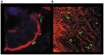
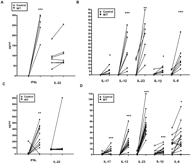
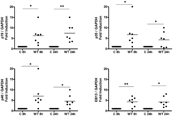
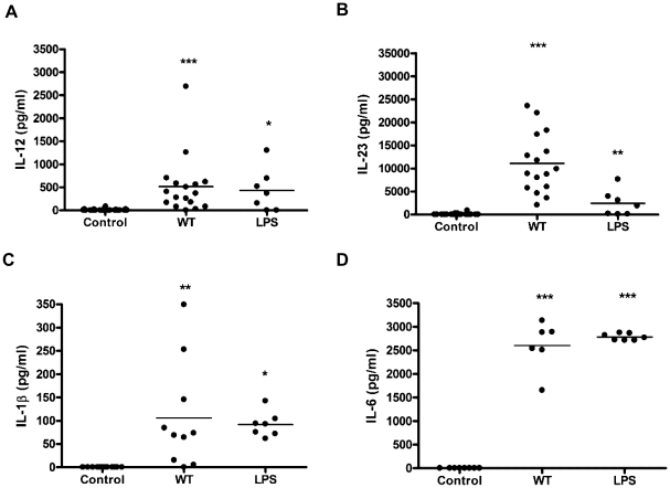
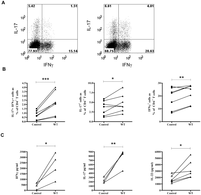
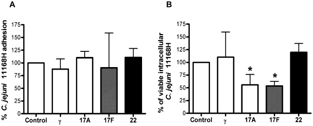
Methodology: Healthy, control paediatric terminal ileum or colonic biopsy tissue was infected with C. jejuni for 8-12 hours. Bacterial colonisation was followed by confocal microscopy and mucosal innate immune responses measured by ELISA. Marked induction of IFNγ with modest increase in IL-22 and IL-17A was noted. Increased mucosal IL-12, IL-23, IL-1β and IL-6 were indicative of a cytokine milieu that may modulate subsequent T-cell mediated immunity. C. jejuni-driven human monocyte-derived dendritic cell activation was followed by analyses of T cell immune responses utilising flow cytometry and ELISA. Significant increase in Th-17, Th-1 and Th-17/Th-1 double-positive cells and corresponding cytokines was observed. The ability of IFNγ, IL-22 and IL-17 cytokines to exert host defence via modulation of C. jejuni adhesion and invasion to intestinal epithelia was measured by standard gentamicin protection assay.

Conclusions: Both innate and adaptive T cell-immunity to C. jejuni infection led to the release of IFNγ, IL-22 and IL-17A; suggesting a critical role for this cytokine triad in establishing host anti-microbial immunity during the acute and effectors phase of infection. In addition, to their known anti-microbial functions; IL-17A and IL-17F reduced the number of intracellular C. jejuni in intestinal epithelia, highlighting a novel aspect of how IL-17 family members may contribute to protective immunity against C. jejuni.


Conflict of interest statement

Competing Interests: The authors have declared that no competing interests exist.

Figures

Figure 1
Figure 1. Ex-vivo colonisation of human intestine by C. jejuni.
Human intestinal biopsies from the terminal ileum were co-cultured for 12 hrs with WT C. jejuni 11168H. Following co-culture, bacteria were localized by immuno-labelling with primary unlabelled anti-campylobacter antibody and secondary FITC labelled antibody (green). Actin filaments including apical brush border were visualized with rhodamine phalloidin (Red). TO-PRO blue was used to counter-stain for nuclei (blue). Whole tissue samples were examined by confocal microscopy. Transverse cross-section of the tissue (a) and an apical view (b) are shown.
Figure 2
Figure 2. Ex-vivo mucosal cytokine responses to C. jejuni 11168H infection.
Paediatric (a) terminal ileum and (b) colonic biopsy tissue were exposed to WT C. jejuni 11168H strain (1×10⁹/ml) bacteria for 8 hours. Post-infection IFNγ and IL-22 (a & c) and IL-17, IL-12, IL-23, IL-1β and IL-6 (b & d) protein were quantified (Data shown with median).
Figure 3
Figure 3. Monocyte-derived DC (DC) cytokine milieu in response to C. jejuni 11168H wild-type strain.
DCs incubated in media alone served as Control (C) or were infected with C. jejuni 11168H wild-type (WT) strain (multiplicity of infection; MOI = 100). mRNA expression of the IL-12 family members (p19, p35, p40, EBI3 at 8 and 24 hours) was quantified by RT-PCR. (a) Gene expression was normalised to GAPDH. Variations in mRNA levels are expressed as fold induction compared to the uninfected control cells. (Median is shown). A representative gel to highlight variation in subunit expression between donors (D) is included (see Figure S1).
Figure 4
Figure 4. C. jejuni 11168H wild-type strain modulates expression of a panel of DC-derived cytokines implicated in human T cell differentiation and survival.
DCs from donors were exposed to WT C. jejuni 11168H wild-type strain or E. coli LPS (10 µg/ml) for 24 hours and cytokine levels measured by ELISA. Significant increase in (a) IL-1β, (b) IL-6, (c) IL-12 and (d) IL-23 protein levels was noted (median is shown).
Figure 5
Figure 5. C. jejuni 11168H infected-DC supernatants promote expansion of Th-17/Th-1 immunity.
PBMCs were enriched for CD4+CD45RO+ memory T cells and stimulated with anti-CD3, anti-CD28 coated beads for 5 days in the presence of supernatants taken from DCs cultured with medium only or with C. jejuni 11168H WT strain. (a) A representative flow cytometric plot of cultured T cells, stimulated with PMA and ionomycin for 3 hours in the presence of Brefeldin A and stained for intracellular IL-17A and IFNγ is shown. (left panel; T cells cultured in uninfected DC supernatants, right panel; T cells cultured in C. jejuni-infected DC supernatants). (b) Number of IL-17+IFNγ+ (left), IL-17+IFNγ (middle) and IFNγ+IL-17 (right) cells as a percentage of CD4+ T cells, as in (a), n = 7. (c) T-cell derived cytokine [IFN-γ (n = 4; left), IL-17 (n = 4; middle) and IL-22 (n = 6; right),] protein quantified 5 days post-stimulation.
Figure 6
Figure 6. IL-17A and IL-17F reduce C. jejuni 11168H intracellular survival in intestinal epithelia.
Confluent Caco-2 cells were exposed to individual cytokines for 24 hours prior to infection with C. jejuni 11168H WT strain (MOI = 100) for 3 hours at 37°C. Cell lysates were serially diluted and plated for total viable bacterial counts (adhesion + invasion). In parallel, another set of infected cells was exposed to 150 µg/ml gentamicin for 2 hours (to kill extracellular adhered bacteria) and lysates plated for enumeration of viable intracellular bacteria. Data represent average percentage cfu obtained in treated versus untreated cells (the latter set at 100%). Statistical analysis of 3 independent experiments performed in duplicate is shown versus untreated cells (Data shown as the median + range).

Similar articles

Cited by

References

    1. Young KT, Davis LM, Dirita VJ. Campylobacter jejuni: molecular biology and pathogenesis. Nat Rev Microbiol. 2007;5:665–679. - PubMed
    1. Poly F, Guerry P. Pathogenesis of Campylobacter. Curr Opin Gastroenterol. 2008;24:27–31. - PubMed
    1. Gradel KO, Nielsen HL, Schonheyder HC, Ejlertsen T, Kristensen B, et al. Increased short- and long-term risk of inflammatory bowel disease after salmonella or campylobacter gastroenteritis. Gastroenterology. 2009;137:495–501. - PubMed
    1. Vucic S, Kiernan MC, Cornblath DR. Guillain-Barre syndrome: An update. J Clin Neurosci. 2009;16:733–741. - PubMed
    1. Oberhelman RA, Gilman RH, Sheen P, Cordova J, Taylor DN, et al. Campylobacter transmission in a Peruvian shantytown: a longitudinal study using strain typing of campylobacter isolates from chickens and humans in household clusters. J Infect Dis. 2003;187:260–269. - PubMed

Publication types

MeSH terms